Advanced 3D cell culture models services for tumor microenvironment
Traditional 2D cell models fail to capture the complexity of human biology in today’s dynamic research environment. Our complex cell model screening CRO-style services leverage Revvity’s advanced imaging technologies and an extensive library of spheroids and primary immune cells, providing more biologically relevant data. This allows for better prediction of human responses to therapeutic interventions in immuno-oncology.
We specialize in generating models for:
- 3D cell culture models: Our advanced screening platforms integrate cell line-derived spheroids and primary immune cell co-cultures, closely replicating in vivo conditions and providing deeper insights into cellular interactions.
- Customized to your requirements: Whether you're focused on assets targeting the cancer cell, or modulating the immune system, we tailor screening assays to match your specific project objectives.
- Leverage precision of gene editing: Engage the power of CRISPR editing in microphysiological systems to explore gene functions and interactions within highly sophisticated biological environments
- Proficiency in advanced biology: Our preclinical services scientists possess solid expertise in working with intricate microphysiological systems, allowing us to provide you with precise and meaningful results.
- Comprehensive solutions: From assay design to high-throughput screening, we provide an end-to-end service that supports every phase of your development, with dedicated project management you can trust.
Learn about our services
T cell infiltration assay
Get valuable insights into the interactions between the immune system and tumors
Understanding how immune cells penetrate and interact with tumors is crucial in providing key insights into the effectiveness of immunotherapies and potential treatment outcomes.
Get valuable insights into the interactions between the immune system and tumors
Understanding how immune cells penetrate and interact with tumors is crucial in providing key insights into the effectiveness of immunotherapies and potential treatment outcomes.
Recreating the tumor microenvironment accurately presents several challenges when establishing a T cell tumor infiltration assay. Challenges include preserving the physiological relevance of immune-tumor interactions in vitro, ensuring optimal co-culture conditions for T cells and tumor cells, and capturing the dynamic infiltration behaviors. Standardization becomes difficult due to variations in tumor architecture and immune cell behavior across different microphysiological systems, while imaging and quantifying T cell infiltration in 3D cell culture models such as spheroids adds further complexity to the process.
What is a T cell infiltration assay?
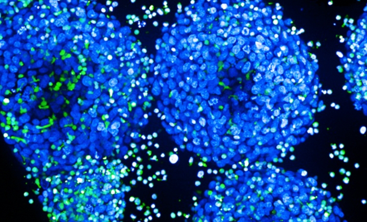
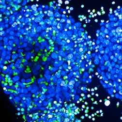
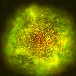
A T cell infiltration assay assesses how T cells can penetrate the 3D structure of an in-vitro tumor tissue, typically in a cancer cell line spheroid model. This assay is valuable for examining the communication between immune cells and tumor cells, especially in immunotherapy, as successful infiltration is essential for the immune system to target and remove cancer cells.
Key readouts at the end of the assay
- Quantifying T Cell infiltration: Measure the number or percentage of T cells that penetrate the tumor mass using Revvity´s high-content imaging hardware and software technologies such as Harmony™ high-content imaging and analysis software.
- Determining T Cell localization: Identify where T cells are situated—whether on the surface, in the tumor periphery, or deep within the tumor tissue—to gain insights into immune cell access and potential resistance mechanisms.
- Assessing cytokine production: Evaluate cytokine levels released by T cells as indicators of immune response and anti-tumor activity, leveraging the performance of HRTF™, AlphaLISA™, and BioLegend LEGENDplex™ technologies.
- Measuring tumor cell viability: Assess tumor cell death or growth inhibition to correlate T cell infiltration with therapeutic effectiveness directly.
T cell killing assay
Unleash the power of immunotherapy with our advanced T cell killing assay
Precise evaluation of cytotoxicity and mechanisms of action using OncoSignature™ cell lines.
Unleash the power of immunotherapy with our advanced T cell killing assay
Precise evaluation of cytotoxicity and mechanisms of action using OncoSignature™ cell lines.
Experience the convenience of outsourcing immuno-oncology drug lead research through Revvity's advanced quantitative 2D and 3D T cell killing platform.
What is a T cell killing assay?
It is a versatile platform for evaluating the efficacy of immuno-oncology therapies. This advanced assay uses techniques to provide microphysiological system readouts, allowing for the comparison of various compounds across both 2D monolayers and 3D spheroid formations of target cells. The T cell killing assay can play a significant role in verifying immunotherapies by offering valuable insights into the cytotoxic functionality of T cells and elucidating compounds´s mode of action by using OncoSignature™ cell lines.
Why choose Revvity's T Cell killing assay service?
- With Revvity's advanced quantitative 2D and 3D T cell killing platform, you can outsource your immuno-oncology drug lead validation with confidence. Our platform provides accurate, reproducible, and physiologically relevant readouts that can help move your research forward.
- Assess the efficacy of your therapeutic candidates by determining the cell death of target cancer spheroids. We use quality effector T cells sourced from diverse human donors to deliver reliable, biologically representative results.
- Revvity preclinical services deliver rapid and relevant imaging readouts along with high-throughput functional data, giving you information to help you make informed decisions faster.
- Leverage our miniaturized in vitro immuno-oncology models to further improve and verify your therapeutic leads. Our models are designed to simulate the tumor-immune interface, providing an accurate reflection of real-world therapeutic performance.
Unraveling the mechanisms behind immunotherapies
Incorporating CRISPR editing into the 3D cell co-culture screening process allows researchers to delve deeper into the mechanisms of action of their compounds. This approach uncovers potential applications beyond the original intent of the therapies, expanding the realm of therapeutic opportunities in immuno-oncology.
Spheroids-based assays for screening
Mimicking tumor structure for more predictive, scalable, high-throughput screening
Replicating tumors in vitro enhances the accuracy of preclinical testing, reducing the risk of costly late-stage failures.
Mimicking tumor structure for more predictive, scalable, high-throughput screening
Replicating tumors in vitro enhances the accuracy of preclinical testing, reducing the risk of costly late-stage failures.
Spheroid-based assays provide a scalable approach for high-throughput screening. By using 3D tumor spheroids in low-attachment plates, simple viability assays, such as Revvity's ATPlite™ 3D, can be easily employed. Spheroid models replicate key features of the tumor microenvironment, offering insights that 2D models often lack.
What are spheroids?
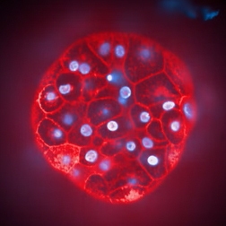
Spheroids are three-dimensional (3D) cell cultures that mimic the architecture of human cell clusters, particularly tumors. Unlike traditional two-dimensional (2D) monolayer cultures, spheroids allow cells to grow in a more natural, 3D structure, providing better cell-to-cell and cell-to-matrix interactions. This makes them highly valuable in cancer research, as they can simulate critical aspects of the tumor microenvironment, such as nutrient gradients, oxygen levels, and drug penetration.
What are the benefits of working with spheroids?
- Increased physiological relevance: Spheroids can more accurately replicate the structure and microenvironment of tumors.
- Enhanced predictive response: The improved physiological relevance of 3D cell culture models can yield more predictable data for in vivo studies.
- Facilitate the simulation of intricate biological processes: Spheroids are better equipped for studying complex biological processes such as tumor growth and metastasis offering deeper insights into disease mechanisms and treatment effects.
- Decrease animal testing: By providing more accurate in vitro models, microphysiological systems can lessen the reliance on animal testing, addressing ethical concerns and reducing the high costs associated.
- Realistic drug penetration and distribution: In spheroids, drug diffusion and distribution can closely mimic in vivo conditions, which is crucial for understanding pharmacokinetics and pharmacodynamics, particularly in solid tumors.
- Applicability for high throughput screening: Advances in automation and imaging technologies have made it possible to use 3D cell culture models in fast and data intensive formats,
- Extended culture duration: 3D cell cultures often allow for longer-term studies, which is essential for assessing chronic toxicity and drug evaluations for several days.
What benefits do you get outsourcing your spheroid project with Revvity’s complex cell model screening services?
Outsourcing your spheroid project with Revvity’s preclinical CRO services means you will receive exceptional support and communication every step of the way. Our dedicated project management team will keep you informed throughout the process, promptly providing clear updates and addressing your needs.
By partnering with us, you'll also gain access to Revvity’s advanced technologies and instrumentation, including our high-content imaging services and powerful analytics software. In addition, we offer multiple protein analytics readouts including BioLegend consumables, enabling multiplexing of your spheroid models. Moreover, you open the opportunity to dive into the genomic drivers of your model with our renowned experience in gene editing and functional genomic screening, to help you to drive your research forward.

Custom complex models project capabilities
Overcoming your complex challenges is what we do
Facing challenges with limited resources or expertise in complex immuno-oncology projects? Collaborate with our specialists to set clear goals, develop a roadmap, and drive breakthrough results.
Overcoming your complex challenges is what we do
Facing challenges with limited resources or expertise in complex immuno-oncology projects? Collaborate with our specialists to set clear goals, develop a roadmap, and drive breakthrough results.
When you work with us, you’ll be updated regularly and frequently on the project´s status as part of our commitment to clear and efficient communication. We will keep you abreast of the project's advancement, achievements, and major changes so you can make well-informed decisions and offer feedback.
You’ll receive a thorough and detailed examination of the outcomes, provided with our dedication to trustworthy data. Our meticulous analytical approaches provide precise and significant analyses, allowing you to decide based on data and shape plans.
Why outsource with Revvity’s preclinical CRO services?
- Boost efficiency: Concentrate on your core strengths while we manage the specialized tasks.
- Expert access: Leverage our global network of skilled professionals for specialized expertise.
- Flexible and scalable: Scale your operations to meet evolving business demands.
- Shared risk: Let us shoulder part of the risk, reducing your operational burden.
- Save time: Focus on strategic initiatives while we handle labor-intensive processes.
Advantages of our complex immuno-oncology cell models projects
- Expert project management: A dedicated team guiding your project from start to finish.
- Innovative assay platform: Miniaturized primary immune cell assays for streamlined testing.
- Tailored donor selection: Customize your studies with specific donor criteria.
- Advanced imaging readouts: Obtain precise and insightful visual data for deeper analysis.
- Proven ImmuSignature™ assays: Rely on the trusted performance of our verified assays.
- Extensive cell line library: Access over 600+ diverse cell lines for comprehensive screening.
Examples of assay development areas
Consult with our scientist to develop your next screening assay, including in the following areas of primary immune cells.
- B cell polarization and suppression assays
- M1 and M2 macrophages polarization
- NK cell Antibody-Dependent and Cell-mediated Cytotoxicity (ADCC) assays
- Antibody-dependent cellular phagocytosis (ADCP)


