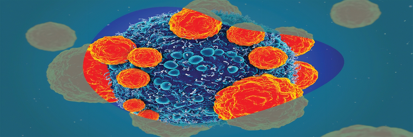
Immunotherapy offers new hope for cancer patients by utilizing the body’s immune system to prevent, control, and eliminate cancer cells. This transformative strategy encompasses various techniques, such as chimeric antigen receptor (CAR) T-cell therapy, immune checkpoint inhibitors (ICIs), monoclonal antibodies, and adoptive cell transfer. While CAR T-cell therapy has shown remarkable success in treating hematological malignancies, its application to solid tumors has faced significant challenges.
The challenge of targeting solid tumors with CAR-T cell therapies
One of the major hurdles of treating solid tumors with CAR T-cell therapies is the unique nature of these cancers. Unlike hematological malignancies, solid tumors exhibit greater heterogeneity and reside within hostile microenvironments with limited blood supply, inadequate oxygen and nutrient delivery, an acidic pH, and a dense extracellular matrix. These factors can impede the effectiveness of CAR T-cells and hinder their ability to infiltrate and destroy tumor cells. There’s also a risk of off-target toxicity, where CAR T-cells may unintentionally target healthy cells expressing the same antigens as tumor cells.
To address these challenges, researchers are turning to innovative methods to model the tumor microenvironment in vitro, providing valuable insights into the effectiveness of CAR T-cell and other immunotherapies.
3D immune cell-killing assays
In the pursuit of advancing our understanding of immune cell function in cancer immunotherapy, 3D immune cell-killing assays have emerged as critical tools. Traditionally, cells have been grown in two-dimensional (2D) cultures on flat surfaces, but more recently, researchers have adopted three-dimensional (3D) cell culture techniques to better mimic the complexities of the in vivo tumor microenvironment.
Various technologies are employed in 3D immune cell-killing assays, each offering unique advantages and insights into immune cell functions and treatment efficacy. These analysis approaches include flow cytometry, high-content analysis, live-cell imaging, and in vivo bioluminescence imaging in small animal models.
However, these 3D assays come with their own set of challenges, including:
- Inconsistent 3D culture growth.
- Difficulty of accurately measuring target cell killing within 3D cultures over time.
- The complexity of reliably measuring immune cell infiltration into 3D cultures.
- Identifying and differentiating between dead cancer cells and dead immune cells.
- Assessing immune cell activity over an adequate duration of time.
- Identifying suitable fluorescent dyes that can penetrate the 3D cell model, do not impair immune cell function, and provide consistent staining over time.
To address these challenges, researchers have developed innovative assays using various analytical technologies to study the antitumor functions of immunotherapies in a 3D environment.


