


Organoids are revolutionizing how we study human development, disease, and drug responses. These three-dimensional, self-assembling cell cultures—derived from stem cells—recapitulate key structural and functional properties of human tissues, bridging the gap between two-dimensional cell cultures and animal models.
Since their development in the early 2010s, organoids have been established for various tissue types, including the liver, intestine, lung, brain, and blood vessels. By faithfully replicating key aspects of tissue architecture and cellular interactions, they can be used to gain unique insights into organ-specific biology. This makes them valuable tools in preclinical research.
The successful culturing of organoids requires specialized knowledge and experience in cell culture techniques, including handling patient-derived samples, maintaining optimal conditions, and monitoring their development over time. Just as crucial is their rigorous characterization, ensuring that they are physiologically relevant, reproducible, and reliable before being used in disease modeling or drug discovery.
A variety of advanced technologies facilitate this characterization process, ensuring findings translate effectively to in vivo human biology. These include:
- Flow cytometry and single-cell RNA sequencing (scRNAseq), which enables precise quantification of cell type diversity
- High-content imaging, which provides detailed insights into tissue architecture
- Multiplex immunoassays, which help assess functional responses at a molecular level
Together, these techniques are essential for ensuring robust and reliable conclusions are drawn from organoid-based experiments.
Organoid characterization techniques.
- Analyzing cellular diversity with flow cytometry
Flow cytometry is a high-throughput technique used to quantify cell diversity within organoids. By utilizing fluorophore-conjugated antibodies, researchers can identify and phenotype various cell types within dissociated organoids.
This method enables the rapid analysis of large cell populations, generating quantitative data to confirm the presence of relevant cell types and assess how well organoids replicate in vivo conditions. Its speed, scalability, and cost-effectiveness make it an essential tool for high-throughput organoid characterization.
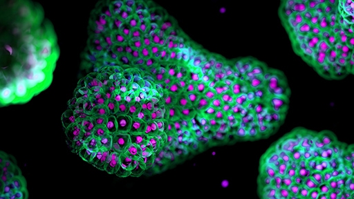
- Understanding organoid morphology with high-content imaging
High-content imaging is an advanced microscopy technique combining automated image acquisition with quantitative analysis, enabling the extraction of multiple cellular parameters from large populations of cells or 3D cell cultures.
This technique provides valuable insights into organoid morphology, including shape, size, organization, and structural remodeling. By capturing changes in tissue architecture over time, high-content imaging helps researchers assess organoid development and detect structural abnormalities in response to perturbations. Standardizing these morphological criteria helps improve the experimental reliability across laboratories, strengthening the quality and impact of organoid studies in both research and therapeutic applications.
- Assessing functional responses with multiplex cytokine immunoassays
Beyond composition and morphology, evaluating organoid function is critical for validating their physiological relevance. Depending on the organoid type, various functions need to be replicated, such as beating (heart organoids), cilia movement (airway organoids), swelling (intestinal organoids), immune responses (gut and lung organoids), or protein secretion (endocrine organoids).
Multiplex immunoassays provide a high-throughput and quantitative way to assess organoid immune function. Compared to traditional ELISAs, which only measure one analyte at a time, multiplex assays allow researchers to simultaneously detect multiple cytokines in a single sample of the culture supernatant. This capability provides a more comprehensive view of the organoid’s functional state, making multiplex assays important tools for studying complex immune interactions in organoid models.
The future of organoid research
As organoid technology continues to evolve, advanced characterization tools will play a pivotal role in standardizing organoid production, enabling more accurate disease modeling and drug screening, and ensuring reproducibility across different laboratories. Tools like flow cytometry, high-content imaging, and multiplex immunoassays will become increasingly crucial for determining organoid composition, morphology, and function.
In addition, the integration of AI and machine learning with these technologies will enable deeper insights into complex organoid datasets, further enhancing their predictive power. To explore real-world applications of these technologies, download our literature review: Advanced characterization tools for organoid research: Analyzing cell type diversity, morphology, and function.


