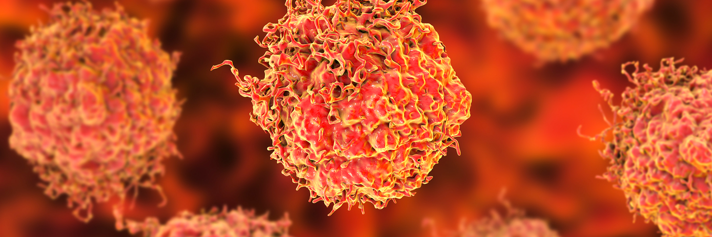
Tumor organoids are becoming increasingly popular advanced model systems for in vitro cancer drug testing. Typically established from patient tumor samples or patient-derived xenografts (PDXs), these 3D cell cultures can replicate the genetic and molecular features of the original tumor. By exposing tumor organoids to various anticancer drugs or drug combinations, researchers can closely monitor their responses, with the aim of identifying the most effective treatment regimen for an individual’s unique tumor biology.
While such an approach holds great promise for personalized medicine, there are often large variations in experimental conditions for organoid culturing and treatment across research groups. This lack of standardization can affect the robustness and reproducibility of results and makes it difficult to compare findings across studies.
Also, most current organoid drug screens use whole-well viability as the sole indicator of effectiveness. This means that important biological information and inter-organoid heterogeneity might be overlooked, both of which are essential for understanding the full impact of treatment.
To address these limitations, researchers at the Erasmus University Medical Center in The Netherlands have optimized the preparation and analysis of prostate cancer organoids for drug testing.1
For their study, the team:
- Identified essential conditions and quality checks to deliver consistent results from their PDX-derived organoids (PDXOs).
- Utilized high-content live-cell imaging to observe any drug-induced effects on tumor growth and cell death at the single-organoid level.
Their work, published in Cells, highlights the power of combining advanced imaging with physiologically relevant 3D organoid models for in vitro cancer drug testing.
Developing an optimized procedure for PDXO drug testing
In the initial stage of their experiment the researchers, led by Annelies Van Hemelryk, set out to optimize the process of preparing prostate cancer PDXOs for drug testing. This involved allowing the organoids to form for 4 to 14 days, then harvesting and filtering them through a fine strainer to reduce size variation. Organoids under 100 μm were then embedded in a hydrogel and seeded in plates at specific densities.
After a four-day recovery period—to ensure they were in a proliferative state—the PDXOs were exposed to various concentrations of the drugs of interest (enzalutamide, docetaxel and cabazitaxel) for 10 days. Following exposure, organoid viability was assessed using a luminescent assay.
This optimized pipeline provided the researchers with reproducible results across replicate experiments, which is crucial for generating reliable data in drug testing.
High-content live-cell imaging of PDXOs
While viability assays—as shown above—are useful for providing a bulk readout of cell viability, they generally lack the ability to distinguish between different drug-induced effects. The researchers therefore developed a live-cell imaging pipeline using the Opera Phenix™ high-content screening system to gain deeper insights into the specific cellular responses induced by drug treatment.
Ten days after drug exposure, the researchers stained living organoids with a combination of Hoechst 33342 nuclear dye and the fluorescent cell death dyes Caspase 3/7 Green and propidium iodide (PI). This multiplex approach allowed them to simultaneously monitor organoid growth along with a range of cell death modalities within individual organoids. The team developed a custom image analysis pipeline to segment and quantify various parameters of individual organoids, including organoid size, number, nuclear counts, and intensities of apoptosis or necrosis markers. The team also implemented quality control measures, such as ensuring a coefficient of variation less than 0.22 and Z-factors greater than 0.4, to help retain the reliability of the imaging results.
Overall, this multiparametric live-cell imaging approach provided the researchers with detailed insights into cellular responses induced by the tested drugs, allowing detection of both cytostatic growth-inhibiting and cytotoxic cell-killing effects within the same assay.
Looking ahead, the researchers suggest that this protocol could be adapted for tumor organoids originating from other cancer types to increase the validity of organoid-based drug testing, and potentially accelerate clinical implementation.
Upscaling your organoid research
At Revvity, we understand the challenges researchers face when scaling up organoid cultures for drug discovery applications. Our guide, ‘Upscaling Organoid Research for Sharper Biological Insights’, has been developed to help you navigate these challenges and find innovative solutions to enhance your organoid research. It covers topics such as optimizing organoid culture conditions, implementing advanced imaging and analysis workflows, and automating organoid handling for high-throughput screening.
For research use only. Not for use in diagnostic procedures.
References:
- Van Hemelryk A, Erkens-Schulze S, Lim L, de Ridder CM, Stuurman DC, Jenster GW, et al. Viability analysis and high-content live-cell imaging for drug testing in prostate cancer xenograft-derived organoids. Cells. 2023 May 12;12(10):1377. doi:10.3390/cells12101377


