
Vega Preclinical Ultrasound System


Vega Preclinical Ultrasound System


Vega Preclinical Ultrasound System


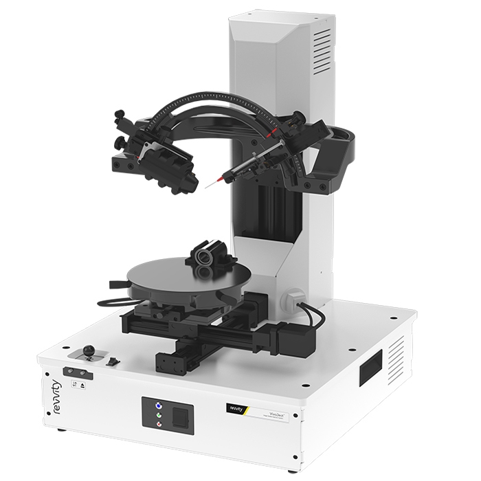
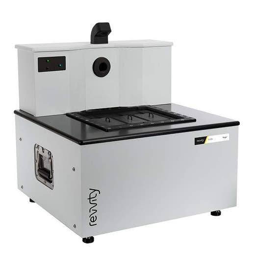
The Vega™ ultrasound system is Revvity's latest addition of leading preclinical in vivo imaging technology. With an innovative design, Vega is a hands-free automated ultrasound platform that delivers high-resolution 2D and 3D imaging in just a few minutes.
Hands-free, automated, high-throughput ultrasound
Product information
Overview
Designed with the researcher in mind, the Vega removes the challenges associated with traditional hand-held ultrasound, and uses a bottom-up imaging approach through the use of automated hands-free transducers located under the imaging stage. This unique design requires minimal training with no dedicated sonographer needed, enables high-throughput imaging, and produces more consistent results than conventional hand-held ultrasound systems.
This powerful ultrasound system gives you:
- Hands-free - Automated transducer positioning and movement
- Easy-to-use requiring minimal training
- High-speed, high-throughput performance with 3 mice scanning in just a few minutes
- 3D widefield acquisitions enabling whole subject imaging
- Standard B-Mode and M-Mode capability
- Shear Wave Elastography (SWE) mode for quantifying tissue stiffness
- Acoustic Angiography (AA) mode for visualization of microvasculature
- Flexible visualization and analysis software
- Fits on the benchtop
Additional product information
Features at a glance
Hands-free
Get more consistent results with a hands-free ultrasound imaging system that removes the challenges associated with conventional hand-held transducers.
Automated
Innovative bottom-up imaging approach through the use of automated transducers located under the imaging stage is easy-to-use and involves minimal training with no dedicated sonographer required.
High-throughput
Acquire scans in less than a minute and increase imaging throughput with 3 mouse sequential scanning and streamlined imaging workflow.
Widefield ultrasound
Get the full picture with fast 3D whole subject imaging for visualizing effects of disease or therapies with broader anatomical context.
Multiple modes for multiple applications
Vega comes standard with two integrated transducers covering many scanning modes for a wide range of applications from high-resolution and vascular imaging to deep tissue imaging, elastography, cardiac imaging, and more.
Elastography
Evaluate and measure tissue stiffness, an important marker used in the clinic to access disease progression, in seconds with Shear Wave Elastography (SWE).
Contrast enhanced ultrasound (CEUS)
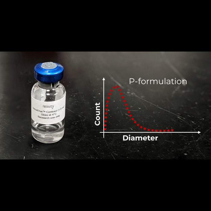
CEUS imaging of microvasculature using Acoustic Angiography mode and VesselVue® microbubble contrast agents to visualize and quantitate tumor vessel network and density or reveal therapy response or injury changes in tissue over time.
Flexible
Through the use of two integrated transducers located under the imaging stage, Vega eliminates the inconvenience of manually swapping out transducers providing ultimate ease and flexibility when switching to different modes.
Customer testimonials

Specifications
| Brand |
Vega
|
|---|---|
| Imaging Modality |
Ultrasound
|
| Unit Size |
1 unit
|
Video gallery

Vega Preclinical Ultrasound System

Vega Preclinical Ultrasound System

Resources
Are you looking for resources, click on the resource type to explore further.
Researchers from Revvity, UNC-Chapel Hill, and NC State University used 3D contrast-enhanced ultrasound imaging technique designed...


How can we help you?
We are here to answer your questions.


