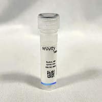
IVISense Arthritis Fluorescent Imaging Panel
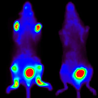
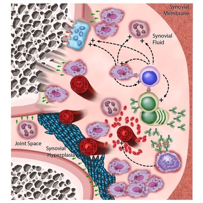
IVISense Arthritis Fluorescent Imaging Panel
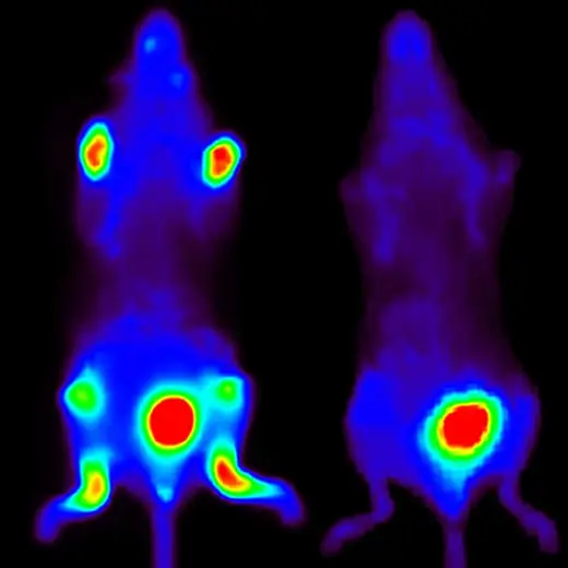
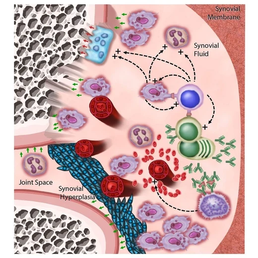


Detect molecular players in arthritis pathology in vivo with Revvity’s IVISense™ Arthritis Fluorescent Imaging Panel.
Standard methods to assess arthritis in animal models typically involve external measurements of limbs such as paw volume, paw thickness, and redness. While these methods determine the overall progression of disease, they do not access the underlying biology associated with the visible effects of the disease. Since arthritis involves not only inflammation pathways but also changes in vasculature and bone morphology, using fluorescent agents that measure each of these changes individually allow for a more complete assessment of the overall clinical pathology.
Revvity' IVISense Arthritis fluorescent panel contains a carefully selected, pre-validated bundle of probes designed for researchers to gain a better understanding of arthritis disease and its progression.
For research use only. Not for use in diagnostic procedures.


IVISense Arthritis Fluorescent Imaging Panel


IVISense Arthritis Fluorescent Imaging Panel


Product information
Overview
The biology of arthritis is complex, involving multiple cellular players, secreted factors, and processes that can lead to sever joint damage. Our fluorescent imaging probes targeting arthritis research allows in vivo imaging of the magnitude of inflammation, bone destruction, cartilage degradation, and vascular leak (edema) in a manner that non-invasively predicts the underlying histopathology of this disease.
The availability of probes at 680 nm and 750 nm wavelengths further offer the opportunity for multiplex imaging of appropriate probe combinations to maximize information gained from research animals.
Additional product information
IVISense Arthritis Fluorescent Imaging Panel includes the following six probes:
| Part Number | Fluorescent Probe | Biological Target |
|---|---|---|
| NEV10003 | IVISense Pan Cathepsin 680 (ProSense 680) | Pan-cathepsin activatable probe: Lysosomal marker of inflammatory cells |
| NEV10011EX | IVISense Vascular 750 (AngeioSense 750EX) | Vascular probe: Leaks into sites of edema |
| NEV10020EX | IVISense Osteo 680 (OsteoSense 680EX) | Bone turnover targeted probe: Detects areas of exposed hydroxyapatite |
| NEV10126 | IVISense MMP 680 (MMPSense 680) | Pan-MMP activatable probe: Secreted marker of inflammatory cells, bone/cartilage degradation |
| NEV11000 | IVISense Cat K 680 FAST | Cathepsin K activatable probe: Secreted marker of osteoclasts, bone degradation |
| NEV11098 | IVISense Cat B 750 FAST | Cathepsin B activatable probe: Lysosomal marker of inflammatory cells |
Specifications
| Brand |
IVISense
|
|---|---|
| Imaging Modality |
Fluorescence
|
| Shipping Conditions |
Shipped in Blue Ice
|
| Therapeutic Area |
Arthritis
|
| Unit Size |
1 kit containing 6 vials
|
Resources
Are you looking for resources, click on the resource type to explore further.
SDS, COAs, Manuals and more
Are you looking for technical documents related to the product? We have categorized them in dedicated sections below. Explore now.
-
Lot number-Lot dateSeptember 11, 2024


How can we help you?
We are here to answer your questions.


