


- Who we serve
- Pharma / Biotech
Revvity Impact Report
Learn how we are increasing our commitment to drive a positive impact on the world.

- Clinical Laboratories
Revvity Impact Report
Learn how we are increasing our commitment to drive a positive impact on the world.
- Healthcare Professionals
Revvity Impact Report
Learn how we are increasing our commitment to drive a positive impact on the world.
- Contract Research Organizations
Revvity Impact Report
Learn how we are increasing our commitment to drive a positive impact on the world.
Revvity Impact Report
Learn how we are increasing our commitment to drive a positive impact on the world.
- Pharma / Biotech
- Products
- 研究・開発
- ゲノム解析
Revvity Impact Report
Learn how we are increasing our commitment to drive a positive impact on the world.
- タンパク質解析
Revvity Impact Report
Learn how we are increasing our commitment to drive a positive impact on the world.
- 細胞分析
Revvity Impact Report
Learn how we are increasing our commitment to drive a positive impact on the world.
- 研究ソリューション
Revvity Impact Report
Learn how we are increasing our commitment to drive a positive impact on the world.
Revvity Impact Report
Learn how we are increasing our commitment to drive a positive impact on the world.
- ゲノム解析
- 臨床・診断
- リプロダクティブ・ヘルス
Revvity Impact Report
Learn how we are increasing our commitment to drive a positive impact on the world.
- 感染症
Revvity Impact Report
Learn how we are increasing our commitment to drive a positive impact on the world.
- がん
Revvity Impact Report
Learn how we are increasing our commitment to drive a positive impact on the world.
- 自己免疫(powered by Euroimmun)
Revvity Impact Report
Learn how we are increasing our commitment to drive a positive impact on the world.
- 内分泌学
Revvity Impact Report
Learn how we are increasing our commitment to drive a positive impact on the world.
- アレルギー
Revvity Impact Report
Learn how we are increasing our commitment to drive a positive impact on the world.
- 神経変性
Revvity Impact Report
Learn how we are increasing our commitment to drive a positive impact on the world.
- ラピッド検査
Revvity Impact Report
Learn how we are increasing our commitment to drive a positive impact on the world.
Revvity Impact Report
Learn how we are increasing our commitment to drive a positive impact on the world.
- リプロダクティブ・ヘルス
- Reagents
Revvity Impact Report
Learn how we are increasing our commitment to drive a positive impact on the world.
- プラットフォームと自動化
- 核酸分離
Revvity Impact Report
Learn how we are increasing our commitment to drive a positive impact on the world.
- 自動液体分注
Revvity Impact Report
Learn how we are increasing our commitment to drive a positive impact on the world.
- 統合ラボオートメーション
Revvity Impact Report
Learn how we are increasing our commitment to drive a positive impact on the world.
- マイクロ流体分析
Revvity Impact Report
Learn how we are increasing our commitment to drive a positive impact on the world.
- 検出システムソリューション
Revvity Impact Report
Learn how we are increasing our commitment to drive a positive impact on the world.
- イメージング
Revvity Impact Report
Learn how we are increasing our commitment to drive a positive impact on the world.
- サンプルのホモジナイゼーション
Revvity Impact Report
Learn how we are increasing our commitment to drive a positive impact on the world.
- IVDプラットフォーム&オートメーション
Revvity Impact Report
Learn how we are increasing our commitment to drive a positive impact on the world.
- 消耗品&アクセサリー
Revvity Impact Report
Learn how we are increasing our commitment to drive a positive impact on the world.
- Instrument Service & Maintenance
Revvity Impact Report
Learn how we are increasing our commitment to drive a positive impact on the world.
Revvity Impact Report
Learn how we are increasing our commitment to drive a positive impact on the world.
- 核酸分離
- Consumables & Accessories
Revvity Impact Report
Learn how we are increasing our commitment to drive a positive impact on the world.
- Signals ソフトウエア
- All Products
Revvity Impact Report
Learn how we are increasing our commitment to drive a positive impact on the world.
- All Solutions
Revvity Impact Report
Learn how we are increasing our commitment to drive a positive impact on the world.
Revvity Impact Report
Learn how we are increasing our commitment to drive a positive impact on the world.
- All Products
- Revvity Omics Services
Revvity Impact Report
Learn how we are increasing our commitment to drive a positive impact on the world.
Revvity Impact Report
Learn how we are increasing our commitment to drive a positive impact on the world.
- 研究・開発
- Services
- Preclinical Services
- Antibody Drug Conjugate Services
Preclinical services
Work with our experienced scientific team and leverage our advanced technologies to help accelerate the preclinical drug discovery process.

- Complex Cell Model Screening Services
Preclinical services
Work with our experienced scientific team and leverage our advanced technologies to help accelerate the preclinical drug discovery process.
- Base Editing Platform
Preclinical services
Work with our experienced scientific team and leverage our advanced technologies to help accelerate the preclinical drug discovery process.
- Immune Cell Screening
Preclinical services
Work with our experienced scientific team and leverage our advanced technologies to help accelerate the preclinical drug discovery process.
- Functional Genomic Screening Services
Preclinical services
Work with our experienced scientific team and leverage our advanced technologies to help accelerate the preclinical drug discovery process.
- Cell Panel Screening
Preclinical services
Work with our experienced scientific team and leverage our advanced technologies to help accelerate the preclinical drug discovery process.
- Cell Line Engineering
Preclinical services
Work with our experienced scientific team and leverage our advanced technologies to help accelerate the preclinical drug discovery process.
- Viral Vector Engineering and Manufacture
Preclinical services
Work with our experienced scientific team and leverage our advanced technologies to help accelerate the preclinical drug discovery process.
Preclinical services
Work with our experienced scientific team and leverage our advanced technologies to help accelerate the preclinical drug discovery process.
- Antibody Drug Conjugate Services
- Revvity Omics Services
- Revvity Omics Clinical Services
T-SPOT.TB testing services.
Revvity's Oxford Diagnostic Laboratories is a large referral laboratory for tuberculosis testing services based on our T-SPOT technology.

- Revvity Omics Pharma Services
T-SPOT.TB testing services.
Revvity's Oxford Diagnostic Laboratories is a large referral laboratory for tuberculosis testing services based on our T-SPOT technology.
T-SPOT.TB testing services.
Revvity's Oxford Diagnostic Laboratories is a large referral laboratory for tuberculosis testing services based on our T-SPOT technology.
- Revvity Omics Clinical Services
- 臨床サービス
- Revvity Omics Clinical Services
T-SPOT.TB testing services.
Revvity's Oxford Diagnostic Laboratories is a large referral laboratory for tuberculosis testing services based on our T-SPOT technology.
- Revvity Omics Pharma Services
T-SPOT.TB testing services.
Revvity's Oxford Diagnostic Laboratories is a large referral laboratory for tuberculosis testing services based on our T-SPOT technology.
- Cellular and Humoral Immunoassays
T-SPOT.TB testing services.
Revvity's Oxford Diagnostic Laboratories is a large referral laboratory for tuberculosis testing services based on our T-SPOT technology.
- Tuberculosis Testing Services
T-SPOT.TB testing services.
Revvity's Oxford Diagnostic Laboratories is a large referral laboratory for tuberculosis testing services based on our T-SPOT technology.
T-SPOT.TB testing services.
Revvity's Oxford Diagnostic Laboratories is a large referral laboratory for tuberculosis testing services based on our T-SPOT technology.
- Revvity Omics Clinical Services
- Customization Services
- Assays and Reagents
T-SPOT.TB testing services.
Revvity's Oxford Diagnostic Laboratories is a large referral laboratory for tuberculosis testing services based on our T-SPOT technology.
- Microplate Services
T-SPOT.TB testing services.
Revvity's Oxford Diagnostic Laboratories is a large referral laboratory for tuberculosis testing services based on our T-SPOT technology.
- Custom Conjugation & Labeling
T-SPOT.TB testing services.
Revvity's Oxford Diagnostic Laboratories is a large referral laboratory for tuberculosis testing services based on our T-SPOT technology.
- Radiosynthesis and Labeling
T-SPOT.TB testing services.
Revvity's Oxford Diagnostic Laboratories is a large referral laboratory for tuberculosis testing services based on our T-SPOT technology.
T-SPOT.TB testing services.
Revvity's Oxford Diagnostic Laboratories is a large referral laboratory for tuberculosis testing services based on our T-SPOT technology.
- Assays and Reagents
- Viral Vector Engineering and Manufacture
- AAV Services
T-SPOT.TB testing services.
Revvity's Oxford Diagnostic Laboratories is a large referral laboratory for tuberculosis testing services based on our T-SPOT technology.
- Lentivirus Services
T-SPOT.TB testing services.
Revvity's Oxford Diagnostic Laboratories is a large referral laboratory for tuberculosis testing services based on our T-SPOT technology.
T-SPOT.TB testing services.
Revvity's Oxford Diagnostic Laboratories is a large referral laboratory for tuberculosis testing services based on our T-SPOT technology.
- AAV Services
- Instrument Service & Maintenance
- AV Services
T-SPOT.TB testing services.
Revvity's Oxford Diagnostic Laboratories is a large referral laboratory for tuberculosis testing services based on our T-SPOT technology.
- Equipment Service Plans
T-SPOT.TB testing services.
Revvity's Oxford Diagnostic Laboratories is a large referral laboratory for tuberculosis testing services based on our T-SPOT technology.
- On-demand Equipment Service
T-SPOT.TB testing services.
Revvity's Oxford Diagnostic Laboratories is a large referral laboratory for tuberculosis testing services based on our T-SPOT technology.
T-SPOT.TB testing services.
Revvity's Oxford Diagnostic Laboratories is a large referral laboratory for tuberculosis testing services based on our T-SPOT technology.
- AV Services
- Customer Training
- Expert-led Training
T-SPOT.TB testing services.
Revvity's Oxford Diagnostic Laboratories is a large referral laboratory for tuberculosis testing services based on our T-SPOT technology.
- Online Training
T-SPOT.TB testing services.
Revvity's Oxford Diagnostic Laboratories is a large referral laboratory for tuberculosis testing services based on our T-SPOT technology.
T-SPOT.TB testing services.
Revvity's Oxford Diagnostic Laboratories is a large referral laboratory for tuberculosis testing services based on our T-SPOT technology.
- Expert-led Training
- OEM ソリューション
T-SPOT.TB testing services.
Revvity's Oxford Diagnostic Laboratories is a large referral laboratory for tuberculosis testing services based on our T-SPOT technology.
T-SPOT.TB testing services.
Revvity's Oxford Diagnostic Laboratories is a large referral laboratory for tuberculosis testing services based on our T-SPOT technology.
- Preclinical Services
- Resources
- Product Support
- Application Support Knowledge base (ASK)
Tech documents, at your fingertips.
Quickly find and download manuals, safety documents, certificates of analysis and more.

- SDS Search
Tech documents, at your fingertips.
Quickly find and download manuals, safety documents, certificates of analysis and more.
- COA/TDS Search
Tech documents, at your fingertips.
Quickly find and download manuals, safety documents, certificates of analysis and more.
- Manual/IFU Search
Tech documents, at your fingertips.
Quickly find and download manuals, safety documents, certificates of analysis and more.
- SpectraViewer
Tech documents, at your fingertips.
Quickly find and download manuals, safety documents, certificates of analysis and more.
- RAD Calculator
Tech documents, at your fingertips.
Quickly find and download manuals, safety documents, certificates of analysis and more.
Tech documents, at your fingertips.
Quickly find and download manuals, safety documents, certificates of analysis and more.
- Application Support Knowledge base (ASK)
- Resource Center
Tech documents, at your fingertips.
Quickly find and download manuals, safety documents, certificates of analysis and more.
- Blog
Tech documents, at your fingertips.
Quickly find and download manuals, safety documents, certificates of analysis and more.
- Events
Tech documents, at your fingertips.
Quickly find and download manuals, safety documents, certificates of analysis and more.
- Customer Training
- Expert-led Training
Tech documents, at your fingertips.
Quickly find and download manuals, safety documents, certificates of analysis and more.
- Online Training
Tech documents, at your fingertips.
Quickly find and download manuals, safety documents, certificates of analysis and more.
Tech documents, at your fingertips.
Quickly find and download manuals, safety documents, certificates of analysis and more.
- Expert-led Training
- Help Center
- Order Support
Tech documents, at your fingertips.
Quickly find and download manuals, safety documents, certificates of analysis and more.
- Contact Us
Tech documents, at your fingertips.
Quickly find and download manuals, safety documents, certificates of analysis and more.
- Technical Support
Tech documents, at your fingertips.
Quickly find and download manuals, safety documents, certificates of analysis and more.
- Instruments Support & Service
Tech documents, at your fingertips.
Quickly find and download manuals, safety documents, certificates of analysis and more.
- SDS Request
Tech documents, at your fingertips.
Quickly find and download manuals, safety documents, certificates of analysis and more.
- COA/TDS Request
Tech documents, at your fingertips.
Quickly find and download manuals, safety documents, certificates of analysis and more.
- Manual/IFU Request
Tech documents, at your fingertips.
Quickly find and download manuals, safety documents, certificates of analysis and more.
- Training Request
Tech documents, at your fingertips.
Quickly find and download manuals, safety documents, certificates of analysis and more.
- Cell Line Terms & Conditions
Tech documents, at your fingertips.
Quickly find and download manuals, safety documents, certificates of analysis and more.
Tech documents, at your fingertips.
Quickly find and download manuals, safety documents, certificates of analysis and more.
- Order Support
- よくあるご質問
Tech documents, at your fingertips.
Quickly find and download manuals, safety documents, certificates of analysis and more.
- Software Downloads
Tech documents, at your fingertips.
Quickly find and download manuals, safety documents, certificates of analysis and more.
- Knowledge Base
- Application support knowledge base (ASK)
Tech documents, at your fingertips.
Quickly find and download manuals, safety documents, certificates of analysis and more.
- Newborn screening disorders
Tech documents, at your fingertips.
Quickly find and download manuals, safety documents, certificates of analysis and more.
- Sample homogenization applications and protocols
Tech documents, at your fingertips.
Quickly find and download manuals, safety documents, certificates of analysis and more.
- TB testing services
Tech documents, at your fingertips.
Quickly find and download manuals, safety documents, certificates of analysis and more.
Tech documents, at your fingertips.
Quickly find and download manuals, safety documents, certificates of analysis and more.
- Application support knowledge base (ASK)
Tech documents, at your fingertips.
Quickly find and download manuals, safety documents, certificates of analysis and more.
- Product Support
Welcome to Revvity: renowned brands and boundless innovation.
Hearing the word "can't" is our call to action!
We help scientists, researchers, and clinicians overcome the world's greatest health obstacles.
View our story
Featured brand: BioLegend
Learn about our world-class antibodies for a diverse set of research areas including immunology, neuroscience, cancer, stem cells and cell biology.
Visit BioLegend.com

JP
Revvity Sites Globally
Select your location.
*e-commerce not available for this region.

HTRF Human Total WRN Detection Kit, 10,000 assay points

HTRF Human Total WRN Detection Kit, 10,000 assay points

WRN Product image

This HTRF kit allows for the cell-based quantitative detection of WRN.
| Feature | Specification |
|---|---|
| Application | Protein Quantification |
| Sample Volume | 16 µL |
This HTRF kit allows for the cell-based quantitative detection of WRN.
Product Variants
Unit Size: 500 assay points
Part #:
64WRNTPEG
Unit Size: 10,000 assay points
Part #:
64WRNTPEH
For research use only. Not for use in diagnostic procedures. All products to be used in accordance with applicable laws and regulations including without limitation, consumption, and disposal requirements under European REACH regulations (EC 1907/2006).
HTRF Human Total WRN Detection Kit, 10,000 assay points
WRN Product image
HTRF Human Total WRN Detection Kit, 10,000 assay points
Product information
Overview
WRN is a protein of the helicase family involved in genome stability-related process including DNA repair, replication, and maintenance via telomere stability. Abnormal WRN functions characterize Werner Syndrome, which is a rare genetic disorder where genome stability is compromised, leading to the accumulation of senescence factors and premature aging in patients. As a disorder related to genome stability and degenerescence, it is also strongly related to increased cancer risks.
Specifications
| Application |
Protein Quantification
|
|---|---|
| Brand |
HTRF
|
| Detection Modality |
HTRF
|
| Lysis Buffer Compatibility |
Lysis Buffer 1
|
| Molecular Modification |
Total
|
| Product Group |
Kit
|
| Sample Volume |
16 µL
|
| Shipping Conditions |
Shipped in Dry Ice
|
| Target |
WRN
|
| Target Class |
Biomarkers
|
| Target Species |
Human
|
| Technology |
TR-FRET
|
| Therapeutic Area |
Inflammation
Oncology
Rare Diseases
|
| Unit Size |
10,000 assay points
|
How it works
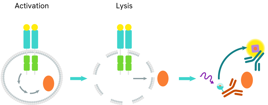
WRN assay principle
The WRN assay measures WRN levels in cells. Unlike Western Blot, the assay is entirely plate-based and does not require gels, electrophoresis, or transfer. The assay uses 2 antibodies, one labeled with a donor fluorophore and the other with an acceptor. In the presence of WRN, the labeled antibodies form an immune complex, bringing the donor fluorophore into close proximity to the acceptor and generating a FRET signal. The signal intensity is directly proportional to the concentration of total protein present in the sample.
WRN two-plate assay protocol
The two-plate protocol involves culturing cells in a 96-well plate before lysis, then transferring lysates into a 384-well low volume detection plate before the addition of WRN HTRF detection reagents. This protocol enables the cells' viability and confluence to be monitored
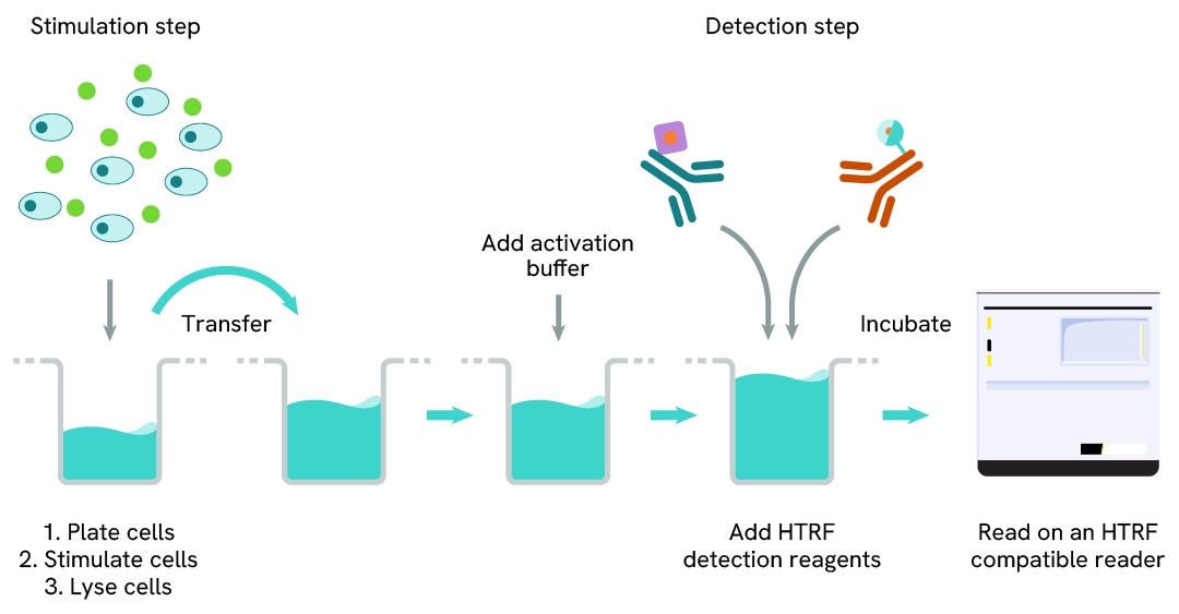
WRN one-plate assay protocol
Detection of WRN with HTRF reagents can be performed in a single plate used for culturing, stimulation, and lysis. No washing steps are required. This HTS designed protocol allows miniaturization while maintaining robust HTRF quality.

Assay validation
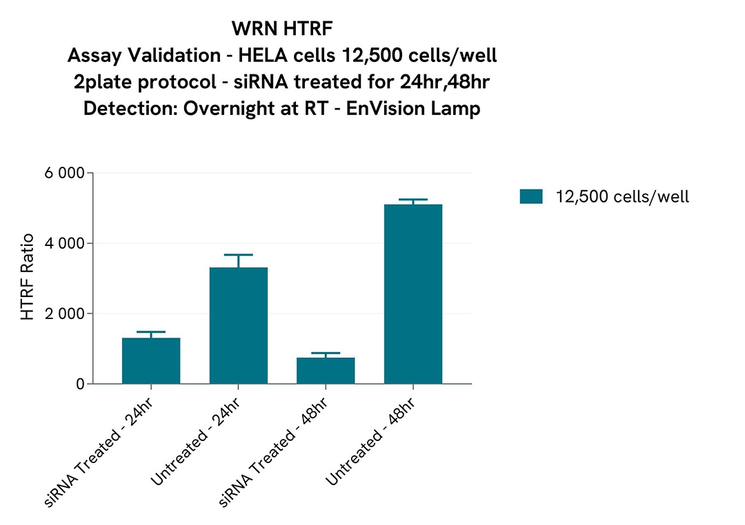
Assay Validation of WRN Detection with siRNA in HeLa cells
HELA cells were plated at 12,500 cells per well under 50 µL in a 96-well plates in complete culture medium. Cells were left to rest for 24hrs before treatment. The initial media was removed and cells were siRNA-treated by adding 50 µl of a mix of Transfection Reagent & siRNA for WRN. Cells were then incubated for 24hr and 48hr at 37°C, 5% CO2.
After incubation, the cells were lysed with 50µL of supplemented lysis buffer #1 at 1X for 30 minutes at RT under gentle shaking, and 16 µL of lysate were transferred into a low volume white microplate before the addition of 2 µL of the HTRF d2 detection reagent and 2 µL HTRF Eu-K detection reagent. The HTRF signal was recorded after ON incubation.
Simplified pathway
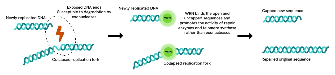
WRN signaling pathway
WRN recruitment takes place at collapsed replication forks of DNA breaks, where it becomes activated by cell cycle kinases like CDKs. There, it stabilizes and supports other helicases like RAD5 which keeps the DNA structure open for repair enzymes, but also downregulates the activity of exonucleases that could otherwise work along the exposed DNA strand and degrade it. This protects the replicated sequence as well as the original one. It also limits the occurrence of degenerated DNA because the genome goes through rounds of replication, thus protecting it from senescence.
If WRN is defective or under expressed, exonucleases are not prevented from degrading replication forks and the genome quickly ages.
Loading...


How can we help you?
We are here to answer your questions.

