


Revvity Sites Globally
Select your location.
*e-commerce not available for this region.

HTRF Human and Mouse CXCL12 Detection Kit, 500 assay points

HTRF Human and Mouse CXCL12 Detection Kit, 500 assay points

CXCL12 Product image

The HTRF CXCL12 kit allows for the simple and rapid quantification of soluble CXCL12 in cell supernatants, providing a fast and no-wash alternative to traditional wash-based ELISA assays. The kit is suitable for the measurement of CXCL12 from both human and mouse cell lines.
| Feature | Specification |
|---|---|
| Application | Protein Quantification |
| Dynamic Range | 93.2 – 8000 pg/mL |
| Limit of Detection | 1.8 pg/mL |
| Limit of Quantification | 7.3 pg/mL |
| Sample Volume | 16 µL |
The HTRF CXCL12 kit allows for the simple and rapid quantification of soluble CXCL12 in cell supernatants, providing a fast and no-wash alternative to traditional wash-based ELISA assays. The kit is suitable for the measurement of CXCL12 from both human and mouse cell lines.
Product Variants
Unit Size: 500 assay points
Part #:
62CXCL12PEG
Unit Size: 10,000 assay points
Part #:
62CXCL12PEH
For research use only. Not for use in diagnostic procedures. All products to be used in accordance with applicable laws and regulations including without limitation, consumption, and disposal requirements under European REACH regulations (EC 1907/2006).
HTRF Human and Mouse CXCL12 Detection Kit, 500 assay points
CXCL12 Product image
HTRF Human and Mouse CXCL12 Detection Kit, 500 assay points
Product information
Overview
CXCL12 (SDF-1) is a homeostatic chemokine constitutively expressed by bone marrow stromal cells, and is present in many other tissues (skin, thymus, lymph nodes, lung, liver). It plays an essential role in the homeostatic regulation of leukocyte traffic, hematopoiesis, organogenesis, cell differentiation, and tissue regeneration.
SDF-1 binds to and activates the CXCR4 receptor. CXCL12 and CXCR4 are overexpressed in various cancer types, and this aberrant expression strongly promotes proliferation, migration, and invasion through multiple signal pathways.
Specifications
| Application |
Protein Quantification
|
|---|---|
| Brand |
HTRF
|
| Detection Modality |
HTRF
|
| Dynamic Range |
93.2 – 8000 pg/mL
|
| Limit of Detection |
1.8 pg/mL
|
| Limit of Quantification |
7.3 pg/mL
|
| Product Group |
Kit
|
| Sample Volume |
16 µL
|
| Shipping Conditions |
Shipped in Dry Ice
|
| Target |
CXCL12
|
| Target Class |
Cytokines
|
| Target Species |
Human
Mouse
|
| Technology |
TR-FRET
|
| Therapeutic Area |
Inflammation
|
| Unit Size |
500 assay points
|
How it works
Principle of the HTRF CXCL12 assay
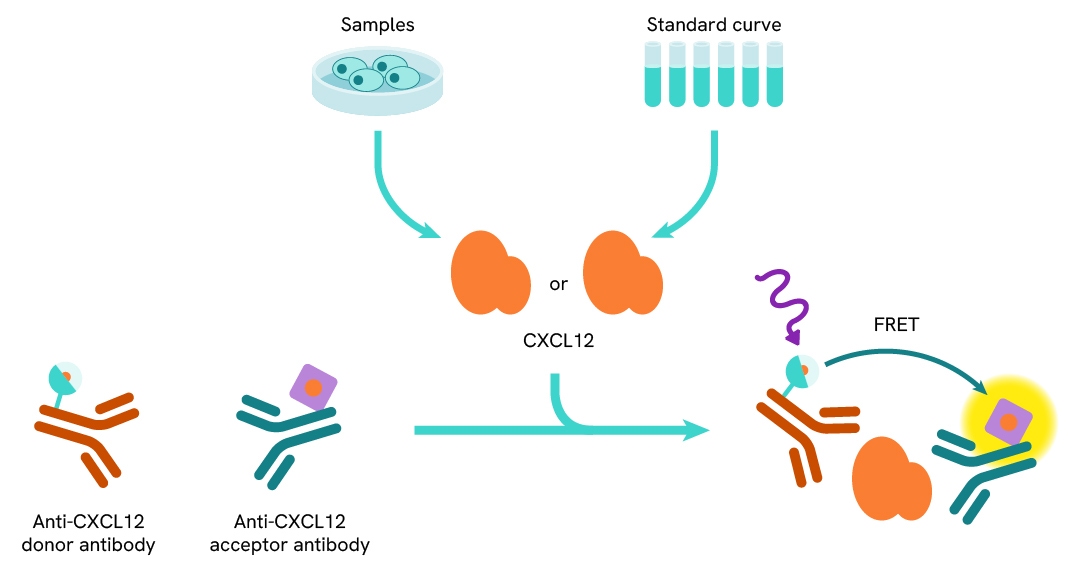
The human and mouse CXCL12 assay is based on a TR-FRET sandwich immunoassay involving two specific antibodies, one labelled with Eu3+ cryptate (donor) and the other with d2 (acceptor). Both antibodies bind to CXCL12, and the donor-acceptor proximity enables a fluorescent TR-FRET signal. The intensity of the signal is directly proportional to the concentration of CXCL12 present in the sample.
Protocol of the HTRF CXCL12 assay
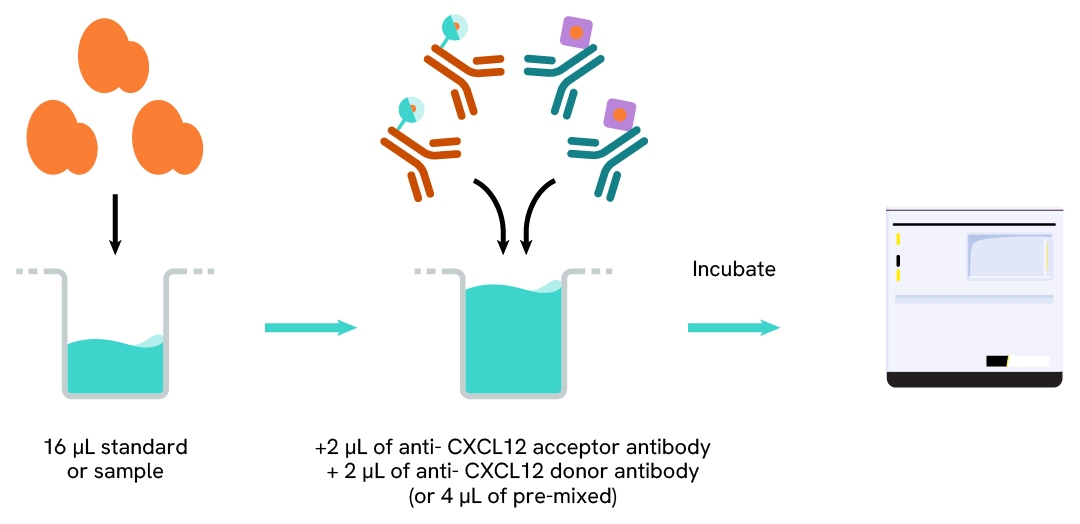
Cell supernatant, sample, or standard is dispensed directly into the assay plate for the detection by HTRF™ reagents (384-well low-volume white plate or Revvity low-volume 96-well plate, in 20 µl). The antibodies labeled with the HTRF donor and acceptor are pre-mixed and added in a single dispensing step, to further streamline the assay procedure. The assay can be run in up to a 1536-well format by simply resizing each addition volume proportionally.
Assay details
| Sample size | 16 µL |
|---|---|
| Final assay volume | 20 µL |
| Kit component | Lyophilized standard, frozen detection antibodies & buffers |
| LOD & LOQ (in Diluent) | 1.8 & 7.3 pg/mL |
| LOD & LOQ (in DMEM) | 18 & 26.3 pg/mL |
| LOD & LOQ (in RMPI) | 14.6 & 24.7 pg/mL |
| Range | 93.2 – 8000 pg/mL |
| Time to result | 4h at RT |
| Species | Human and mouse |
Analytical performance
Intra-assay precision table
Each of the 3 samples was measured 24 times, and the % CV was calculated for each sample.
Samples were mouse recombinant CXCL12.
| Sample | Mean [CXCL12] pg/mL | CV |
|---|---|---|
| 1 | 110 | 2% |
| 2 | 514 | 4% |
| 3 | 1983 | 5% |
| Mean CV | 4% |
Inter-assay precision table
Each of the samples was measured in 3 independent experiments (3 days), and the % CV was calculated for each sample. Samples were mouse recombinant CXCL12.
| Sample | Mean [CXCL12] pg/mL | CV |
|---|---|---|
| 1 | 211 | 3% |
| 2 | 641 | 3% |
| 3 | 2132 | 3% |
| Mean CV | 3% |
Dilution linearity
The excellent recovery percentages obtained from these experiments show the good dilution linearity of the assay. Samples were mouse recombinant CXCL12 serially diluted in diluent 5.
| Dilution factor | Expected CXCL12 concentration (pg/mL) | Measured CXCL12 concentration (pg/mL) | % dilution recovery |
|---|---|---|---|
| neat | - | 5381 |
- |
| 2 | 2690 | 2617 | 97% |
| 4 | 1354 | 1421 | 105% |
| 8 | 672 | 716 | 106% |
| 16 | 336 | 359 | 106% |
| 32 | 168 | 176 | 104% |
| Mean | 103% | ||
Cross-reactivity
Cross reactivities were assessed using recombinant proteins and tested at 1000 pg/mL. Signals were interpolated on the assay standard curve to check concentrations. The assay is human CXCL12 isoform α specific, as isoforms b and γ were not detected. The assay is also compatible with mouse CXCL12.
| Tested protein | Cross reactivity |
|---|---|
| Human CXCL12 isoform α | 100% |
| Human CXCL12 isoform b | 0% |
| Human CXCL12 isoform γ | 0% |
| Mouse CXCL12 | 100% |
Assay validation
Validation of HTRF CXCL12 detection on human cell supernatants
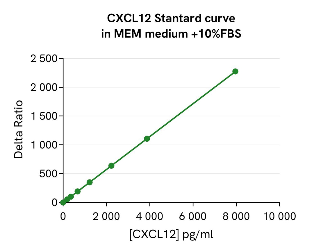
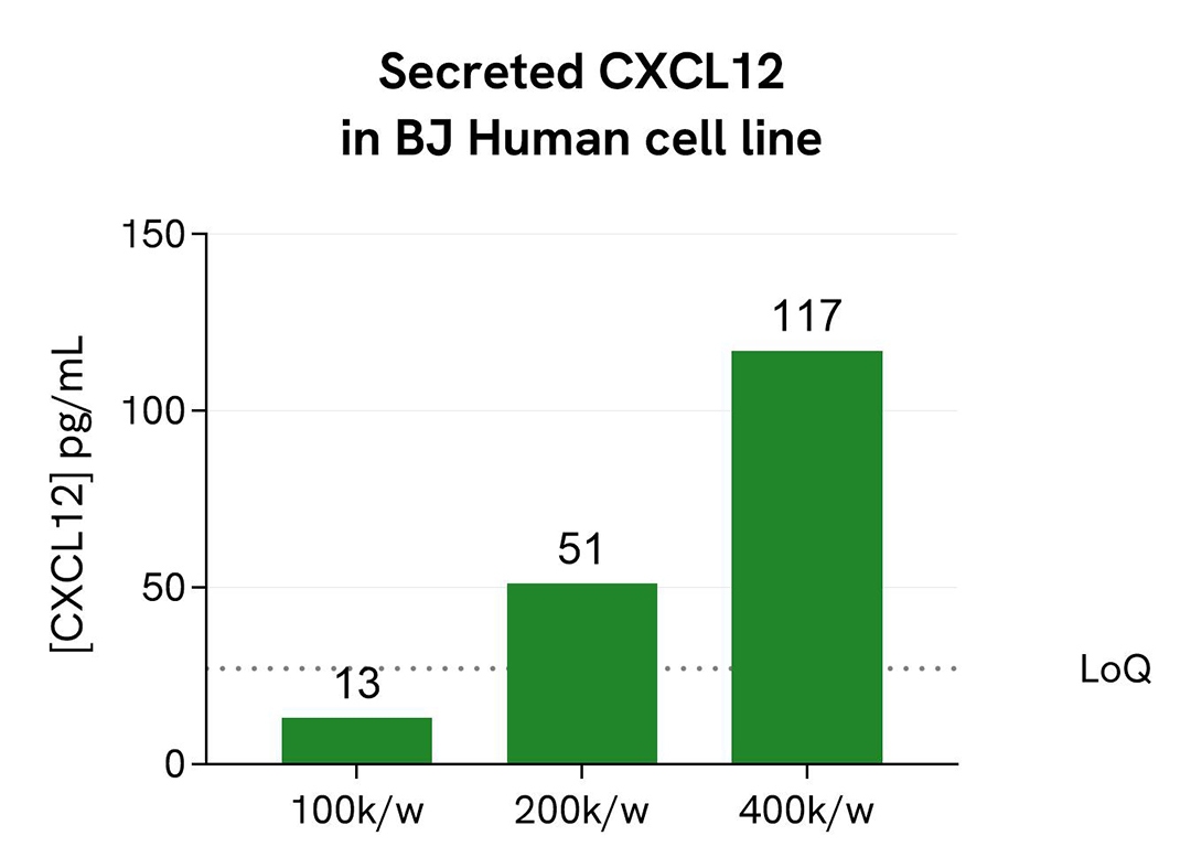
BJ human fibroblast cells were plated in a 96-well culture plate at 100k, 200k, and 4000k cells/well in MEM medium with 10% FBS for 24H at 37°C. Then, 16 µL of supernatants were transferred into a white detection plate (384 wells, low volume) and 4 µL of the HTRF CXCL12 detection reagents were added. The HTRF signal was recorded after 4h of incubation at room temperature.
The standard curve prepared in MEM medium + 10% FBS enabled the interpolation of the CXCL12 concentration in each sample.
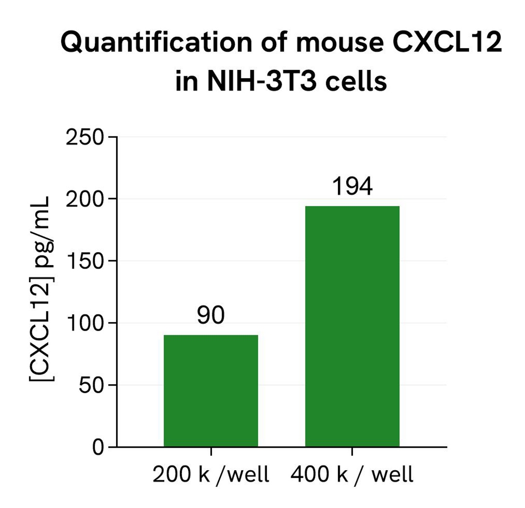
Validation of HTRF CXCL12 detection on mouse cell supernatants
NIH-3T3 mouse fibroblast cells were plated in a 96-well culture plate at 200k and 400k cells/well in DMEM medium with 10% FBS for 24H at 37°C. Then, 16 µL of supernatants were transferred into a white detection plate (384 wells, low volume) and 4 µL of the HTRF CXCL12 detection reagents were added. The HTRF signal was recorded after 4h of incubation at room temperature.
The standard curve prepared in DMEM medium + 10% FBS enabled the interpolation of the CXCL12 concentration in each sample.
Loading...


How can we help you?
We are here to answer your questions.

