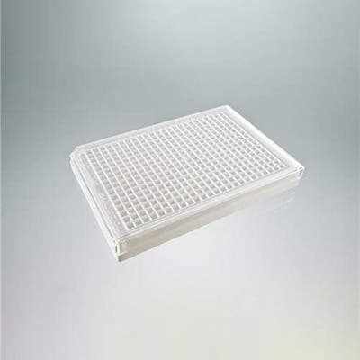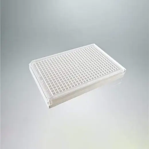
CytoStar-T 384-well plates, 5 plates

CytoStar-T 384-well plates, 5 plates


384-well clear-bottom, scintillating microplate for cell-based radiometric proximity assays.
| Feature | Specification |
|---|---|
| Surface Treatment |
Scintillant Sterile TC-treated |
| Color | White |
| Detection Modality | Radiometric |
| Lids Included? | Yes |
| Material | Polystyrene |
| Sterility | Sterile |
| Plate Format | 384 wells |
| Well Shape | Flat-bottom |
| Well Volume | 105 µL |
| Working Volume | 25 - 90 µL |
384-well clear-bottom, scintillating microplate for cell-based radiometric proximity assays.


CytoStar-T 384-well plates, 5 plates


CytoStar-T 384-well plates, 5 plates


Product information
Overview
The CytoStar-T 96-well scintillating microplate is treated for the growth of tissue culture cells. The microplates are made sterile by gamma-irradiation and treated for adherence of cells by plasma discharge which renders the cell attachment surface hydrophilic. The tissue culture surface is not a coating but rather a stably modified surface with random functional groups covalently bound. The lid has condensation rings that reduce the risk of well-to-well contamination and at the same time reduces evaporation from individual wells, minimizing the potential for "edge effects".
CytoStar-T™ scintillating microplates are designed for non-invasive quantitation in real-time analysis of a wide spectrum of biological reactions in cells under normal physiological conditions. Analyses include cell adhesion, cell signaling (e.g., receptor-ligand binding), cell motility, cell proliferation, normal cellular metabolism, metabolite transport, as well as drug processing (intake and efflux). They are sterile, tissue culture treated microplates designed not only for the growth of adherent but also suspension cell cultures (after plate centrifugation or cells settle). The integral, planar, transparent base of each well is composed of a proprietary homogeneous mixture of scintillants and polystyrene. The transparent nature of the base permits the observation of growth of cells plated in the well. Furthermore, radioisotopes having suitable decay characteristics (3H, 14C, 35S, 33P, 45Ca, 125I), brought into proximity with the scintillant contained within the base by virtue of the biological processes within the cells, will have that radioactive decay converted to a light signal. This blue light signal can then be detected and quantified using a PMT-based radiometric detection instrument. The amount of light generated is proportional to the amount of radioisotope within, or associated with the cells. Unit size 100 plates per case.
The CytoStar-T 384-well scintillating microplate is treated for the growth of tissue culture cells. The microplates are made sterile by gamma-irradiation and treated for adherence of cells by plasma discharge which renders the cell attachment surface hydrophilic. The tissue culture surface is not a coating but rather a stably modified surface with random functional groups covalently bound. The lid has condensation rings that reduce the risk of well-to-well contamination and at the same time reduces evaporation from individual wells, minimizing the potential for "edge effects".
CytoStar-T™ scintillating microplates are designed for non-invasive quantitation in real-time analysis of a wide spectrum of biological reactions in cells under normal physiological conditions. Analyses include cell adhesion, cell signaling (e.g., receptor-ligand binding), cell motility, cell proliferation, normal cellular metabolism, metabolite transport, as well as drug processing (intake and efflux). They are sterile, tissue culture treated microplates designed not only for the growth of adherent but also suspension cell cultures (after plate centrifugation or cells settle). The integral, planar, transparent base of each well is composed of a proprietary homogeneous mixture of scintillants and polystyrene. The transparent nature of the base permits the observation of growth of cells plated in the well. Furthermore, radioisotopes having suitable decay characteristics (3H, 14C, 35S, 33P, 45Ca, 125I), brought into proximity with the scintillant contained within the base by virtue of the biological processes within the cells, will have that radioactive decay converted to a light signal. This blue light signal can then be detected and quantified using a PMT-based radiometric detection instrument. The amount of light generated is proportional to the amount of radioisotope within, or associated with the cells. Unit size 5 plates per case.
Specifications
| Color |
White
|
|---|---|
| Plate Format |
384 wells
|
| Well Shape |
Flat-bottom
|
| Well Volume |
105 µL
|
| Working Volume |
25 - 90 µL
|
| Application |
Cell Culture
Radiometric
Research
|
|---|---|
| Automation Compatible |
Yes
|
| Brand |
CytoStar-T
|
| Detection Modality |
Radiometric
|
| Format |
384-well Microplate
|
| Instrument Compatibility |
MicroBeta
|
| Lids Included? |
Yes
|
| Material |
Polystyrene
|
| Shipping Conditions |
Shipped Ambient
|
| Sterility |
Sterile
|
| Surface Treatment |
Scintillant
Sterile
TC-treated
|
| Unit Size |
5 per case
|
Resources
Are you looking for resources, click on the resource type to explore further.


How can we help you?
We are here to answer your questions.


