


Revvity Sites Globally
Select your location.
*e-commerce not available for this region.

AlphaLISA SureFire Ultra Human & Mouse Total IRF5 Detection Kit, 100 Assay Points


AlphaLISA SureFire Ultra Human & Mouse Total IRF5 Detection Kit, 100 Assay Points


AlphaLISA Surefire Ultra Total Protein


The AlphaLISA™ SureFire® Ultra™ Human and Mouse Total IRF5 assay is a sandwich immunoassay for quantitative detection of total IRF5 in cellular lysates using Alpha Technology.
| Feature | Specification |
|---|---|
| Application | Cell Signaling |
| Sample Volume | 30 µL |
The AlphaLISA™ SureFire® Ultra™ Human and Mouse Total IRF5 assay is a sandwich immunoassay for quantitative detection of total IRF5 in cellular lysates using Alpha Technology.
Product Variants
Unit Size: 100 assay points
Part #:
ALSU-TIRF5-A-HV
Unit Size: 500 assay points
Part #:
ALSU-TIRF5-A500
Unit Size: 10,000 assay points
Part #:
ALSU-TIRF5-A10K
Unit Size: 50,000 assay points
Part #:
ALSU-TIRF5-A50K
For research use only. Not for use in diagnostic procedures. All products to be used in accordance with applicable laws and regulations including without limitation, consumption, and disposal requirements under European REACH regulations (EC 1907/2006).
AlphaLISA SureFire Ultra Human & Mouse Total IRF5 Detection Kit, 100 Assay Points
AlphaLISA Surefire Ultra Total Protein
AlphaLISA SureFire Ultra Human & Mouse Total IRF5 Detection Kit, 100 Assay Points
Product information
Overview
Interferon regulator factor 5, IRF5, is a transcriptional regulator of type I interferon (IFN-alpha and IFN-beta)-dependent immune responses. This transcription factor plays a key role in the innate immune response against DNA and RNA viruses. IRF5 is present in the cytoplasm of uninfected cells in an inactive form. Various mediators of the pathways that follow viral infection can overexpress and phosphorylate IRF5. IRF5 is associated with various cancers and autoimmune diseases.
The AlphaLISA SureFire Ultra Human and Mouse Total IRF5 Detection Kit is a sandwich immunoassay for the quantitative detection of total IRF5 in cellular lysates, using Alpha Technology.
Formats:
- The HV (high volume) kit contains reagents to run 100 wells in 96-well format, using a 60 μL reaction volume.
- The 500-point kit contains enough reagents to run 500 wells in 384-well format, using a 20 μL reaction volume.
- The 10,000-point kit contains enough reagents to run 10,000 wells in 384-well format, using a 20 μL reaction volume.
- The 50,000-point kit contains enough reagents to run 50,000 wells in 384-well format, using a 20 μL reaction volume.
AlphaLISA SureFire Ultra kits are compatible with:
- Cell and tissue lysates
- Antibody modulators
- Biotherapeutic antibodies
Alpha SureFire kits can be used for:
- Cellular kinase assays
- Receptor activation studies
- Screening
Specifications
| Application |
Cell Signaling
|
|---|---|
| Automation Compatible |
Yes
|
| Brand |
AlphaLISA SureFire Ultra
|
| Detection Modality |
Alpha
|
| Lysis Buffer Compatibility |
Lysis Buffer
|
| Molecular Modification |
Total
|
| Product Group |
Kit
|
| Sample Volume |
30 µL
|
| Shipping Conditions |
Shipped in Blue Ice
|
| Target |
IRF5
|
| Target Class |
Phosphoproteins
|
| Target Species |
Human
Mouse
|
| Technology |
Alpha
|
| Therapeutic Area |
Inflammation
Oncology
|
| Unit Size |
100 assay points
|
Video gallery
AlphaLISA SureFire Ultra Human & Mouse Total IRF5 Detection Kit, 100 Assay Points
AlphaLISA SureFire Ultra Human & Mouse Total IRF5 Detection Kit, 100 Assay Points
How it works
Total-AlphaLISA SureFire Ultra assay principle
The Total-AlphaLISA SureFire Ultra assay measures the expression level of a protein target in a cell lysate.
The Total-AlphaLISA SureFire Ultra assay uses two antibodies which recognize two different distal epitopes on the targeted protein. AlphaLISA assays require two bead types: Acceptor and Donor beads. Acceptor beads are coated with a proprietary CaptSure™ agent to specifically immobilize the assay specific antibody, labeled with a CaptSure™ tag. Donor beads are coated with streptavidin to capture one of the detection antibodies, which is biotinylated. In the presence of targeted protein, the two antibodies bring the Donor and Acceptor beads in close proximity whereby the singlet oxygen transfers energy to excite the Acceptor bead, allowing the generation of a luminescent Alpha signal. The amount of light emission is directly proportional to the quantity of protein present in the sample.
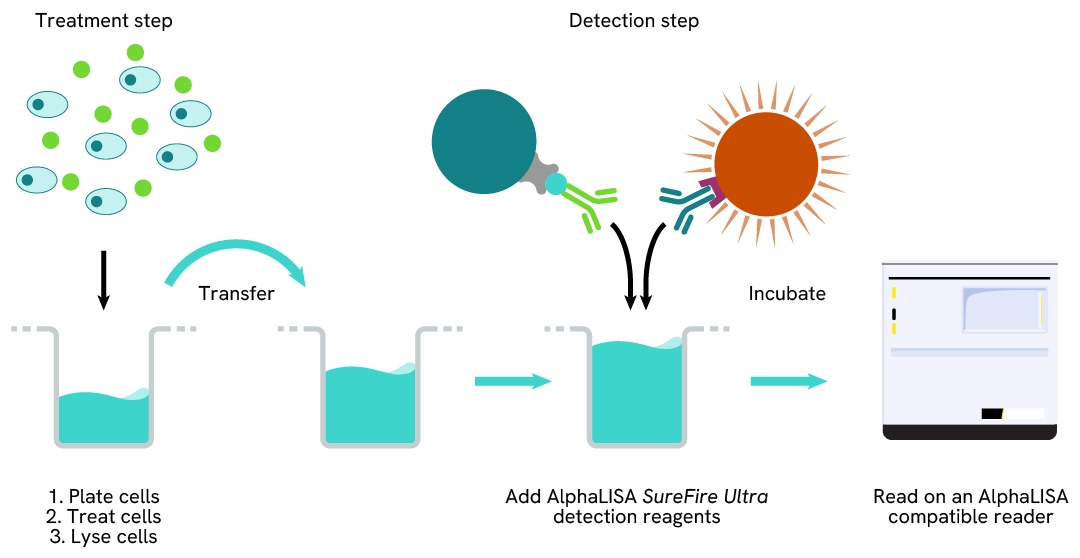
Total-AlphaLISA SureFire Ultra two-plate assay protocol
The two-plate protocol involves culturing and treating the cells in a 96-well plate before lysis, then transferring lysates into a 384-well OptiPlate™ plate before the addition of Total-AlphaLISA SureFire Ultra detection reagents. This protocol permits the cells' viability and confluence to be monitored. In addition, lysates from a single well can be used to measure multiple targets.
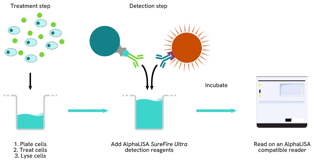
Total-AlphaLISA SureFire Ultra one-plate assay protocol
Detection of Total target protein with AlphaLISA SureFire Ultra reagents can be performed in a single plate used for culturing, treatment, and lysis. No washing steps are required. This HTS designed protocol allows for miniaturization while maintaining AlphaLISA SureFire Ultra quality.
Assay validation
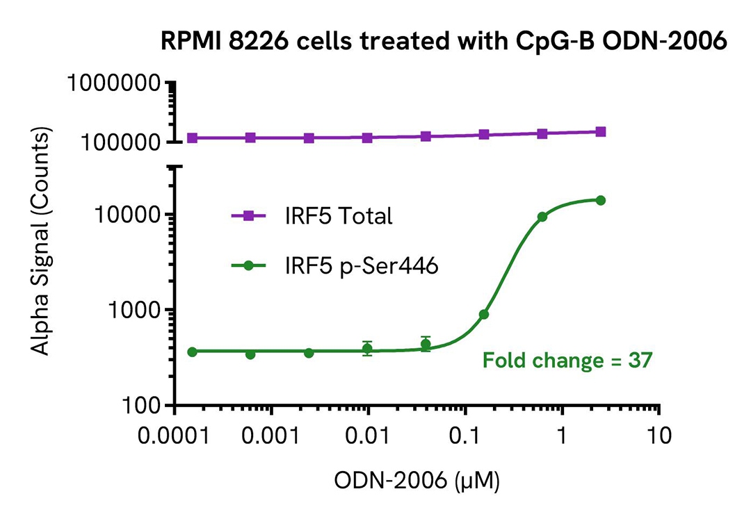
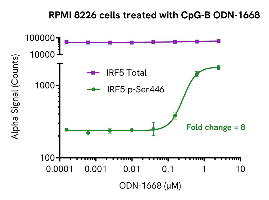
Validation of Phospho (Ser446)/Total IRF5 in CpG-B treated cells
RPMI 8226 cells were seeded in a 96-well plate (500,000 cells/well) in complete medium and incubated for approximately 10 minutes at 37°C, 5% CO2. The cells were treated with increasing concentrations of CpG-B, ODN-2006 or ODN-1668 (MedChem Express, HY-150218, HY-150726 respectively) for 6 hours.
After treatment, the cells were spun down at 1200 rpm for 5 minutes, washed with HBSS + 0.1 % BSA and then lysed with 100 µL of Lysis Buffer for 10 minutes at RT with shaking (350 rpm). IRF5 Phospho (Ser446) and Total levels were evaluated using respective AlphaLISA SureFire Ultra assays. For the detection step, 10 µL of cell lysate (approximately 50,000 cells for ODN-2006 experiment and 20,000 cells for ODN-1668 experiment) were transferred into a 384-well white OptiPlate, followed by 5 µL of Acceptor mix and incubated for 1 hour at RT. Finally, 5 µL of Donor mix was then added to each well and incubated for 1 hour at RT in the dark. The plate was read on an Envision using standard AlphaLISA settings.
As expected, CpG-B triggered a dose-dependent increase in the levels Phospho (Ser446) while Total IRF5 levels remained unchanged.
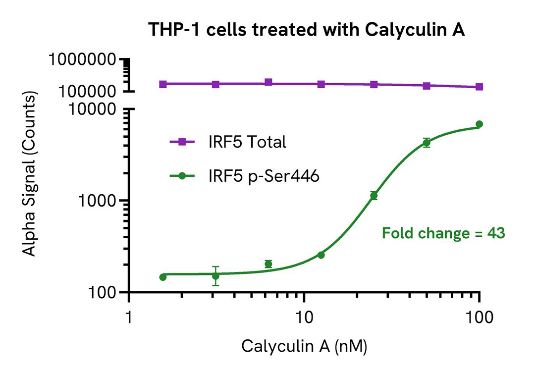
Validation of Phospho (Ser446)/Total IRF5 in Calyculin A treated cells
THP-1 cells were seeded in a 96-well plate (40,000 cells/well) in complete medium and incubated for approximately 10 minutes at 37°C, 5% CO2. The cells were treated with increasing concentrations of Calyculin A for 2.5 hours.
After treatment, the cells were spun down at 1200 rpm for 5 minutes, washed with HBSS + 0.1 % BSA and lysed with 100 µL of Lysis Buffer for 10 minutes at RT with shaking (350 rpm). IRF5 Phospho (Ser446) and Total levels were evaluated using respective AlphaLISA SureFire Ultra assays. For the detection step, 10 µL of cell lysate (approximately 4,000 cells) were transferred into a 384-well white OptiPlate, followed by 5 µL of Acceptor mix and incubated for 1 hour at RT. Finally, 5 µL of Donor mix was then added to each well and incubated for 1 hour at RT in the dark. The plate was read on an Envision using standard AlphaLISA settings.
As expected, Calyculin A triggered a dose-dependent increase in the levels IRF5 Phospho (Ser446) while Total IRF5 levels remained unchanged.
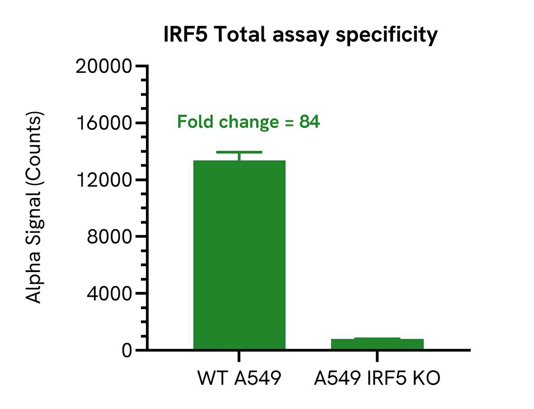
Specificity of IRF5 Total assay
Total IRF5 protein levels were assessed in A549 wild type (WT) and A549 IRF5 knockout (KO) (Abcam, ab301006) cells. Both cell lines were grown to confluency in a T75 flask in medium at 37°C, 5% CO2. Each flask was lysed in 2 mL of Lysis Buffer for 10 minutes at RT with shaking (350 rpm). Lysates were then evaluated for Total IRF5 using the AlphaLISA SureFire Ultra kit.
For the detection step, 10 µL of cell lysate was transferred into a 384-well white OptiPlate, followed by 5 µL of Acceptor Mix and incubated for 1 hour at RT. Finally, 5 µL of Donor Mix was added to each well and incubated for 1 hour at RT in the dark. The plate was read on an Envision using standard AlphaLISA settings.
As expected, Total IRF5 protein was detected only in A549 WT cells and no signal was detected in the KO IRF5 A549 cell line, demonstrating assay specificity.
Assay versatility
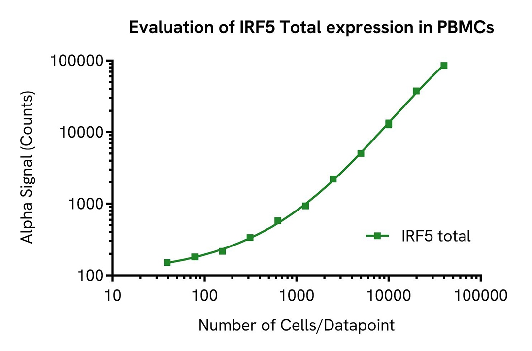
Evaluation of IRF5 Total expression in Peripheral Blood Mononuclear Cells
Peripheral Blood Mononuclear Cells (PBMCs) were isolated from healthy donors using Ficoll® Plaque Plus (Merck GE17-1440-02).
Cells were seeded in 96-well plate (400,000 cells/well) and lysed in 100 µL of Lysis Buffer for 10 minutes at RT with shaking (350 rpm). Generated lysate was then serially diluted in Lysis Buffer and evaluated using the AlphaLISA SureFire Ultra Total IRF5. For the detection step, 10 µL of cell lysate (starting from approximately 40,000 cells) were transferred into a 384-well white OptiPlate, followed by 5 µL of Acceptor mix and incubated for 1 hour at RT. Finally, 5 µL of Donor mix was then added to each well and incubated for 1 hour at RT in the dark. The plate was read on an Envision using standard AlphaLISA settings. The number of cells/datapoint is indicated on graph.
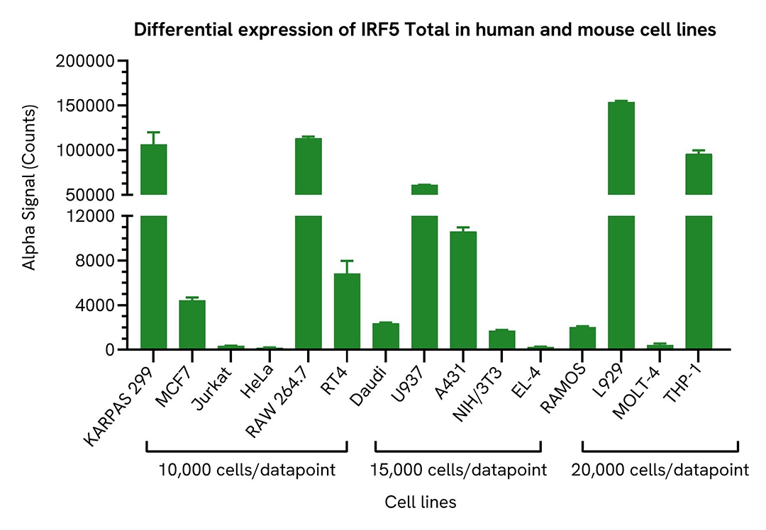
Differential expression of IRF5 Total in various cell lines
Human and mouse cell lysates were diluted with Lysis Buffer. Approximate number of cells/datapoint is indicated on graph. Total IRF5 levels were evaluated using the AlphaLISA SureFira Ultra assay kit. For the detection step, 10 µL of cell lysate was transferred into a 384-well white OptiPlate, followed by 5 µL of Acceptor Mix and incubated for 1 hour at RT. Finally, 5 µL of Donor Mix was then added to each well and incubated for 1 hour at RT in the dark. The plate was read on an Envision using standard AlphaLISA settings.
Total IRF5 expression varies depending upon cell type. As expected, a very high level of expression was detected in human cell lines: THP-1, KARPAS-299 and U937 and mouse cell lines: RAW 264.7 and L929.
Resources
Are you looking for resources, click on the resource type to explore further.
Brochure
Alpha SureFire Ultra no-wash immunoassay catalog
Discover Alpha SureFire® Ultra™ assays, the no-wash cellular kinase assays leveraging Revvity's exclusive bead-based technology...
Brochure
Species compatibility for HTRF, AlphaLISA SureFire Ultra and Alpha SureFire Ultra Multiplex assays
This document includes detailed tables listing HTRF™, AlphaLISA™ SureFire® Ultra™, and Alpha SureFire® Ultra™ Multiplex assays...
Loading...


How can we help you?
We are here to answer your questions.

