


- Who we serve
- Pharma / Biotech
Revvity Impact Report
Learn how we are increasing our commitment to drive a positive impact on the world.

- Clinical Laboratories
Revvity Impact Report
Learn how we are increasing our commitment to drive a positive impact on the world.
- Healthcare Professionals
Revvity Impact Report
Learn how we are increasing our commitment to drive a positive impact on the world.
- Contract Research Organizations
Revvity Impact Report
Learn how we are increasing our commitment to drive a positive impact on the world.
Revvity Impact Report
Learn how we are increasing our commitment to drive a positive impact on the world.
- Pharma / Biotech
- Products
- 研究・開発
- ゲノム解析
Revvity Impact Report
Learn how we are increasing our commitment to drive a positive impact on the world.
- タンパク質解析
Revvity Impact Report
Learn how we are increasing our commitment to drive a positive impact on the world.
- 細胞分析
Revvity Impact Report
Learn how we are increasing our commitment to drive a positive impact on the world.
- 研究ソリューション
Revvity Impact Report
Learn how we are increasing our commitment to drive a positive impact on the world.
Revvity Impact Report
Learn how we are increasing our commitment to drive a positive impact on the world.
- ゲノム解析
- 臨床・診断
- リプロダクティブ・ヘルス
Revvity Impact Report
Learn how we are increasing our commitment to drive a positive impact on the world.
- 感染症
Revvity Impact Report
Learn how we are increasing our commitment to drive a positive impact on the world.
- がん
Revvity Impact Report
Learn how we are increasing our commitment to drive a positive impact on the world.
- 自己免疫(powered by Euroimmun)
Revvity Impact Report
Learn how we are increasing our commitment to drive a positive impact on the world.
- 内分泌学
Revvity Impact Report
Learn how we are increasing our commitment to drive a positive impact on the world.
- アレルギー
Revvity Impact Report
Learn how we are increasing our commitment to drive a positive impact on the world.
- 神経変性
Revvity Impact Report
Learn how we are increasing our commitment to drive a positive impact on the world.
- ラピッド検査
Revvity Impact Report
Learn how we are increasing our commitment to drive a positive impact on the world.
Revvity Impact Report
Learn how we are increasing our commitment to drive a positive impact on the world.
- リプロダクティブ・ヘルス
- Reagents
Revvity Impact Report
Learn how we are increasing our commitment to drive a positive impact on the world.
- プラットフォームと自動化
- 核酸分離
Revvity Impact Report
Learn how we are increasing our commitment to drive a positive impact on the world.
- 自動液体分注
Revvity Impact Report
Learn how we are increasing our commitment to drive a positive impact on the world.
- 統合ラボオートメーション
Revvity Impact Report
Learn how we are increasing our commitment to drive a positive impact on the world.
- マイクロ流体分析
Revvity Impact Report
Learn how we are increasing our commitment to drive a positive impact on the world.
- 検出システムソリューション
Revvity Impact Report
Learn how we are increasing our commitment to drive a positive impact on the world.
- イメージング
Revvity Impact Report
Learn how we are increasing our commitment to drive a positive impact on the world.
- サンプルのホモジナイゼーション
Revvity Impact Report
Learn how we are increasing our commitment to drive a positive impact on the world.
- IVDプラットフォーム&オートメーション
Revvity Impact Report
Learn how we are increasing our commitment to drive a positive impact on the world.
- 消耗品&アクセサリー
Revvity Impact Report
Learn how we are increasing our commitment to drive a positive impact on the world.
- Instrument Service & Maintenance
Revvity Impact Report
Learn how we are increasing our commitment to drive a positive impact on the world.
Revvity Impact Report
Learn how we are increasing our commitment to drive a positive impact on the world.
- 核酸分離
- Consumables & Accessories
Revvity Impact Report
Learn how we are increasing our commitment to drive a positive impact on the world.
- Signals ソフトウエア
- All Products
Revvity Impact Report
Learn how we are increasing our commitment to drive a positive impact on the world.
- All Solutions
Revvity Impact Report
Learn how we are increasing our commitment to drive a positive impact on the world.
Revvity Impact Report
Learn how we are increasing our commitment to drive a positive impact on the world.
- All Products
- Revvity Omics Services
Revvity Impact Report
Learn how we are increasing our commitment to drive a positive impact on the world.
Revvity Impact Report
Learn how we are increasing our commitment to drive a positive impact on the world.
- 研究・開発
- Services
- Preclinical Services
- Antibody Drug Conjugate Services
Preclinical services
Work with our experienced scientific team and leverage our advanced technologies to help accelerate the preclinical drug discovery process.

- Complex Cell Model Screening Services
Preclinical services
Work with our experienced scientific team and leverage our advanced technologies to help accelerate the preclinical drug discovery process.
- Base Editing Platform
Preclinical services
Work with our experienced scientific team and leverage our advanced technologies to help accelerate the preclinical drug discovery process.
- Immune Cell Screening
Preclinical services
Work with our experienced scientific team and leverage our advanced technologies to help accelerate the preclinical drug discovery process.
- Functional Genomic Screening Services
Preclinical services
Work with our experienced scientific team and leverage our advanced technologies to help accelerate the preclinical drug discovery process.
- Cell Panel Screening
Preclinical services
Work with our experienced scientific team and leverage our advanced technologies to help accelerate the preclinical drug discovery process.
- Cell Line Engineering
Preclinical services
Work with our experienced scientific team and leverage our advanced technologies to help accelerate the preclinical drug discovery process.
- Viral Vector Engineering and Manufacture
Preclinical services
Work with our experienced scientific team and leverage our advanced technologies to help accelerate the preclinical drug discovery process.
Preclinical services
Work with our experienced scientific team and leverage our advanced technologies to help accelerate the preclinical drug discovery process.
- Antibody Drug Conjugate Services
- Revvity Omics Services
- Revvity Omics Clinical Services
T-SPOT.TB testing services.
Revvity's Oxford Diagnostic Laboratories is a large referral laboratory for tuberculosis testing services based on our T-SPOT technology.

- Revvity Omics Pharma Services
T-SPOT.TB testing services.
Revvity's Oxford Diagnostic Laboratories is a large referral laboratory for tuberculosis testing services based on our T-SPOT technology.
T-SPOT.TB testing services.
Revvity's Oxford Diagnostic Laboratories is a large referral laboratory for tuberculosis testing services based on our T-SPOT technology.
- Revvity Omics Clinical Services
- 臨床サービス
- Revvity Omics Clinical Services
T-SPOT.TB testing services.
Revvity's Oxford Diagnostic Laboratories is a large referral laboratory for tuberculosis testing services based on our T-SPOT technology.
- Revvity Omics Pharma Services
T-SPOT.TB testing services.
Revvity's Oxford Diagnostic Laboratories is a large referral laboratory for tuberculosis testing services based on our T-SPOT technology.
- Cellular and Humoral Immunoassays
T-SPOT.TB testing services.
Revvity's Oxford Diagnostic Laboratories is a large referral laboratory for tuberculosis testing services based on our T-SPOT technology.
- Tuberculosis Testing Services
T-SPOT.TB testing services.
Revvity's Oxford Diagnostic Laboratories is a large referral laboratory for tuberculosis testing services based on our T-SPOT technology.
T-SPOT.TB testing services.
Revvity's Oxford Diagnostic Laboratories is a large referral laboratory for tuberculosis testing services based on our T-SPOT technology.
- Revvity Omics Clinical Services
- Customization Services
- Assays and Reagents
T-SPOT.TB testing services.
Revvity's Oxford Diagnostic Laboratories is a large referral laboratory for tuberculosis testing services based on our T-SPOT technology.
- Microplate Services
T-SPOT.TB testing services.
Revvity's Oxford Diagnostic Laboratories is a large referral laboratory for tuberculosis testing services based on our T-SPOT technology.
- Custom Conjugation & Labeling
T-SPOT.TB testing services.
Revvity's Oxford Diagnostic Laboratories is a large referral laboratory for tuberculosis testing services based on our T-SPOT technology.
- Radiosynthesis and Labeling
T-SPOT.TB testing services.
Revvity's Oxford Diagnostic Laboratories is a large referral laboratory for tuberculosis testing services based on our T-SPOT technology.
T-SPOT.TB testing services.
Revvity's Oxford Diagnostic Laboratories is a large referral laboratory for tuberculosis testing services based on our T-SPOT technology.
- Assays and Reagents
- Viral Vector Engineering and Manufacture
- AAV Services
T-SPOT.TB testing services.
Revvity's Oxford Diagnostic Laboratories is a large referral laboratory for tuberculosis testing services based on our T-SPOT technology.
- Lentivirus Services
T-SPOT.TB testing services.
Revvity's Oxford Diagnostic Laboratories is a large referral laboratory for tuberculosis testing services based on our T-SPOT technology.
T-SPOT.TB testing services.
Revvity's Oxford Diagnostic Laboratories is a large referral laboratory for tuberculosis testing services based on our T-SPOT technology.
- AAV Services
- Instrument Service & Maintenance
- AV Services
T-SPOT.TB testing services.
Revvity's Oxford Diagnostic Laboratories is a large referral laboratory for tuberculosis testing services based on our T-SPOT technology.
- Equipment Service Plans
T-SPOT.TB testing services.
Revvity's Oxford Diagnostic Laboratories is a large referral laboratory for tuberculosis testing services based on our T-SPOT technology.
- On-demand Equipment Service
T-SPOT.TB testing services.
Revvity's Oxford Diagnostic Laboratories is a large referral laboratory for tuberculosis testing services based on our T-SPOT technology.
T-SPOT.TB testing services.
Revvity's Oxford Diagnostic Laboratories is a large referral laboratory for tuberculosis testing services based on our T-SPOT technology.
- AV Services
- Customer Training
- Expert-led Training
T-SPOT.TB testing services.
Revvity's Oxford Diagnostic Laboratories is a large referral laboratory for tuberculosis testing services based on our T-SPOT technology.
- Online Training
T-SPOT.TB testing services.
Revvity's Oxford Diagnostic Laboratories is a large referral laboratory for tuberculosis testing services based on our T-SPOT technology.
T-SPOT.TB testing services.
Revvity's Oxford Diagnostic Laboratories is a large referral laboratory for tuberculosis testing services based on our T-SPOT technology.
- Expert-led Training
- OEM ソリューション
T-SPOT.TB testing services.
Revvity's Oxford Diagnostic Laboratories is a large referral laboratory for tuberculosis testing services based on our T-SPOT technology.
T-SPOT.TB testing services.
Revvity's Oxford Diagnostic Laboratories is a large referral laboratory for tuberculosis testing services based on our T-SPOT technology.
- Preclinical Services
- Resources
- Product Support
- Application Support Knowledge base (ASK)
Tech documents, at your fingertips.
Quickly find and download manuals, safety documents, certificates of analysis and more.

- SDS Search
Tech documents, at your fingertips.
Quickly find and download manuals, safety documents, certificates of analysis and more.
- COA/TDS Search
Tech documents, at your fingertips.
Quickly find and download manuals, safety documents, certificates of analysis and more.
- Manual/IFU Search
Tech documents, at your fingertips.
Quickly find and download manuals, safety documents, certificates of analysis and more.
- SpectraViewer
Tech documents, at your fingertips.
Quickly find and download manuals, safety documents, certificates of analysis and more.
- RAD Calculator
Tech documents, at your fingertips.
Quickly find and download manuals, safety documents, certificates of analysis and more.
Tech documents, at your fingertips.
Quickly find and download manuals, safety documents, certificates of analysis and more.
- Application Support Knowledge base (ASK)
- Resource Center
Tech documents, at your fingertips.
Quickly find and download manuals, safety documents, certificates of analysis and more.
- Blog
Tech documents, at your fingertips.
Quickly find and download manuals, safety documents, certificates of analysis and more.
- Events
Tech documents, at your fingertips.
Quickly find and download manuals, safety documents, certificates of analysis and more.
- Customer Training
- Expert-led Training
Tech documents, at your fingertips.
Quickly find and download manuals, safety documents, certificates of analysis and more.
- Online Training
Tech documents, at your fingertips.
Quickly find and download manuals, safety documents, certificates of analysis and more.
Tech documents, at your fingertips.
Quickly find and download manuals, safety documents, certificates of analysis and more.
- Expert-led Training
- Help Center
- Order Support
Tech documents, at your fingertips.
Quickly find and download manuals, safety documents, certificates of analysis and more.
- Contact Us
Tech documents, at your fingertips.
Quickly find and download manuals, safety documents, certificates of analysis and more.
- Technical Support
Tech documents, at your fingertips.
Quickly find and download manuals, safety documents, certificates of analysis and more.
- Instruments Support & Service
Tech documents, at your fingertips.
Quickly find and download manuals, safety documents, certificates of analysis and more.
- SDS Request
Tech documents, at your fingertips.
Quickly find and download manuals, safety documents, certificates of analysis and more.
- COA/TDS Request
Tech documents, at your fingertips.
Quickly find and download manuals, safety documents, certificates of analysis and more.
- Manual/IFU Request
Tech documents, at your fingertips.
Quickly find and download manuals, safety documents, certificates of analysis and more.
- Training Request
Tech documents, at your fingertips.
Quickly find and download manuals, safety documents, certificates of analysis and more.
- Cell Line Terms & Conditions
Tech documents, at your fingertips.
Quickly find and download manuals, safety documents, certificates of analysis and more.
Tech documents, at your fingertips.
Quickly find and download manuals, safety documents, certificates of analysis and more.
- Order Support
- よくあるご質問
Tech documents, at your fingertips.
Quickly find and download manuals, safety documents, certificates of analysis and more.
- Software Downloads
Tech documents, at your fingertips.
Quickly find and download manuals, safety documents, certificates of analysis and more.
- Knowledge Base
- Application support knowledge base (ASK)
Tech documents, at your fingertips.
Quickly find and download manuals, safety documents, certificates of analysis and more.
- Newborn screening disorders
Tech documents, at your fingertips.
Quickly find and download manuals, safety documents, certificates of analysis and more.
- Sample homogenization applications and protocols
Tech documents, at your fingertips.
Quickly find and download manuals, safety documents, certificates of analysis and more.
- TB testing services
Tech documents, at your fingertips.
Quickly find and download manuals, safety documents, certificates of analysis and more.
Tech documents, at your fingertips.
Quickly find and download manuals, safety documents, certificates of analysis and more.
- Application support knowledge base (ASK)
Tech documents, at your fingertips.
Quickly find and download manuals, safety documents, certificates of analysis and more.
- Product Support
Welcome to Revvity: renowned brands and boundless innovation.
Hearing the word "can't" is our call to action!
We help scientists, researchers, and clinicians overcome the world's greatest health obstacles.
View our story
Featured brand: BioLegend
Learn about our world-class antibodies for a diverse set of research areas including immunology, neuroscience, cancer, stem cells and cell biology.
Visit BioLegend.com

JP
Revvity Sites Globally
Select your location.
*e-commerce not available for this region.

AlphaLISA SureFire Ultra Human Phospho-AKT1/2/3 (Ser473) Detection Kit, 50,000 Assay Points

 View All
AlphaLISA SureFire Ultra Human Phospho-AKT1/2/3 (Ser473) Detection Kit, 50,000 Assay Points


AlphaLISA SureFire Ultra Phospho-Protein


The AlphaLISA™ SureFire® Ultra™ p-AKT1/2/3 (Ser473) assay is a sandwich immunoassay for quantitative detection of phospho-AKT1/2/3 (phosphorylated on Ser473) in cellular lysates using Alpha Technology.
| Feature | Specification |
|---|---|
| Application | Cell Signaling |
| Sample Volume | 10 µL |
The AlphaLISA™ SureFire® Ultra™ p-AKT1/2/3 (Ser473) assay is a sandwich immunoassay for quantitative detection of phospho-AKT1/2/3 (phosphorylated on Ser473) in cellular lysates using Alpha Technology.
Product Variants
Unit Size: 500 assay points
Part #:
ALSU-PAKT-B500
Unit Size: 10,000 assay points
Part #:
ALSU-PAKT-B10K
Unit Size: 50,000 assay points
Part #:
ALSU-PAKT-B50K
Unit Size: 100 assay points
Part #:
ALSU-PAKT-B-HV
For research use only. Not for use in diagnostic procedures. All products to be used in accordance with applicable laws and regulations including without limitation, consumption, and disposal requirements under European REACH regulations (EC 1907/2006).
AlphaLISA SureFire Ultra Human Phospho-AKT1/2/3 (Ser473) Detection Kit, 50,000 Assay Points
AlphaLISA SureFire Ultra Phospho-Protein
AlphaLISA SureFire Ultra Human Phospho-AKT1/2/3 (Ser473) Detection Kit, 50,000 Assay Points
Supporting products you might need
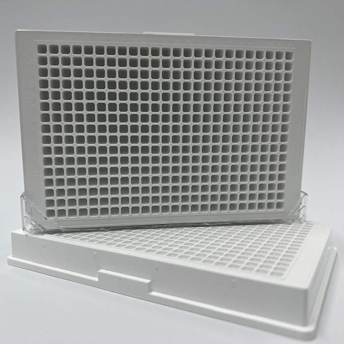
Don't forget your microplates
Part #: 6007290
Part #: 6007299
Product information
Overview
The AKT serine/threonine kinase, also known as protein kinase B (PKB), is a crucial component of the PI3K/AKT/mTOR signaling pathway. In humans, there are three AKT isoforms (AKT1, AKT2, and AKT3) encoded by separate genes. AKT plays a central role in various cellular processes, including cell survival, proliferation, metabolism, and angiogenesis. Activation of AKT occurs through phosphorylation at two key sites: Thr308 and Ser473. The phosphorylation of Ser473 is particularly important for full activation of AKT and its downstream signaling. AKT signaling is frequently dysregulated in various diseases, especially cancer, where hyperactivation of the pathway contributes to tumor growth, metastasis, and therapy resistance.
The AlphaLISA SureFire Ultra Human Phospho-AKT1/2/3 (Ser473) Detection Kit is a sandwich immunoassay designed for the quantitative detection of phosphorylated AKT (Ser473) in cellular lysates, using Alpha technology
Formats
- The HV (high volume) kit contains reagents to run 100 wells in 96-well format, using a 60 µL reaction volume.
- The 500-point kit contains enough reagents to run 500 wells in 384-well format, using a 20 µL reaction volume.
- The 10,000-point kit contains enough reagents to run 10,000 wells in 384-well format, using a 20 µL reaction volume.
- The 50,000-point kit contains enough reagents to run 50,000 wells in 384-well format, using a 20 µL reaction volume.
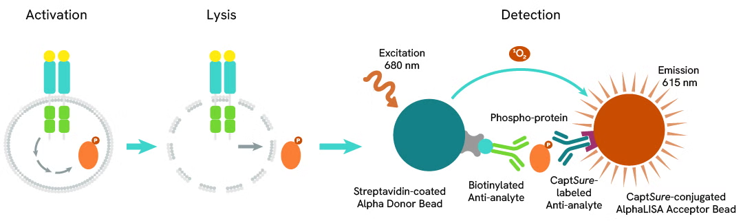
In the AlphaLISA™ SureFire® Ultra™ assay, Donor beads are coated with streptavidin to capture one of the antibodies, which is biotinylated. Acceptor beads are coated with a proprietary CaptSure™ agent that immobilizes the other antibody, labeled with a CaptSure™ tag. In the presence of phosphorylated protein, the two antibodies bring the Donor and Acceptor beads close together, generating signal. The amount of light emission is directly proportional to the amount of phosphoprotein present in the sample.
AlphaLISA SureFire Ultra kits are compatible with
- Cell and tissue lysates
- Antibody modulators
- Biotherapeutic antibodies
Alpha SureFire kits can be used for
- Cellular kinase assays
- Receptor activation studies
- Screening
Specifications
| Application |
Cell Signaling
|
|---|---|
| Automation Compatible |
Yes
|
| Brand |
AlphaLISA SureFire Ultra
|
| Cellular or Signaling Pathway |
PI3K/Akt/mTOR
|
| Detection Modality |
Alpha
|
| Lysis Buffer Compatibility |
Lysis Buffer
|
| Molecular Modification |
Phosphorylation
|
| Product Group |
Kit
|
| Sample Volume |
10 µL
|
| Shipping Conditions |
Shipped in Blue Ice
|
| Target |
Akt1/2/3
|
| Target Class |
Phosphoproteins
|
| Target Species |
Human
|
| Technology |
Alpha
|
| Therapeutic Area |
Cardiovascular
Central Nervous System
Metabolic
|
| Unit Size |
50,000 assay points
|
Image gallery
AlphaLISA SureFire Ultra Human Phospho-AKT1/2/3 (Ser473) Detection Kit, 50,000 Assay Points
AlphaLISA SureFire Ultra Human Phospho-AKT1/2/3 (Ser473) Detection Kit, 50,000 Assay Points
Video gallery
AlphaLISA SureFire Ultra Human Phospho-AKT1/2/3 (Ser473) Detection Kit, 50,000 Assay Points
AlphaLISA SureFire Ultra Human Phospho-AKT1/2/3 (Ser473) Detection Kit, 50,000 Assay Points
Citations
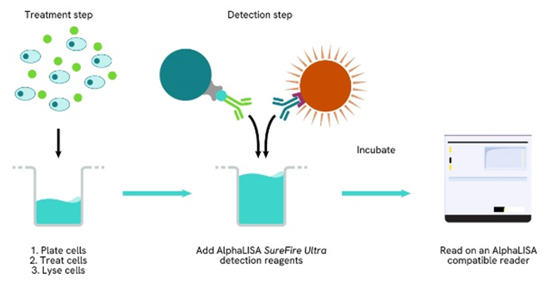
How it works
Phospho-AlphaLISA SureFire Ultra assay principle
The Phospho-AlphaLISA SureFire Ultra assay measures a protein target when phosphorylated at a specific residue.
The assay uses two antibodies which recognize the phospho epitope and a distal epitope on the targeted protein. AlphaLISA assays require two bead types: Acceptor and Donor beads. Acceptor beads are coated with a proprietary CaptSure™ agent to specifically immobilize the assay specific antibody, labeled with a CaptSure™ tag. Donor beads are coated with streptavidin to capture one of the detection antibodies, which is biotinylated. In the presence of phosphorylated protein, the two antibodies bring the Donor and Acceptor beads in close proximity whereby the singlet oxygen transfers energy to excite the Acceptor bead, allowing the generation of a luminescent Alpha signal. The amount of light emission is directly proportional to the quantity of phosphoprotein present in the sample.
Phospho-AlphaLISA SureFire Ultra two-plate assay protocol
The two-plate protocol involves culturing and treating the cells in a 96-well plate before lysis, then transferring lysates into a 384-well OptiPlate™ plate before the addition of Phospho-AlphaLISA SureFire Ultra detection reagents. This protocol permits the cells' viability and confluence to be monitored. In addition, lysates from a single well can be used to measure multiple targets.
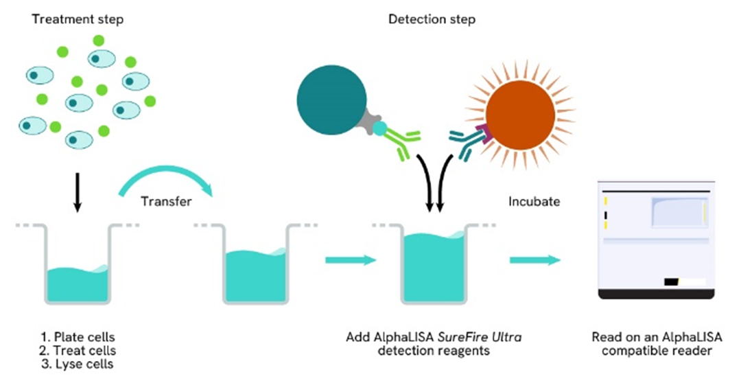
Phospho-AlphaLISA SureFire Ultra one-plate assay protocol
Detection of Phosphorylated target protein with AlphaLISA SureFire Ultra reagents can be performed in a single plate used for culturing, treatment, and lysis. No washing steps are required. This HTS designed protocol allows for miniaturization while maintaining AlphaLISA SureFire Ultra quality.
Assay validation
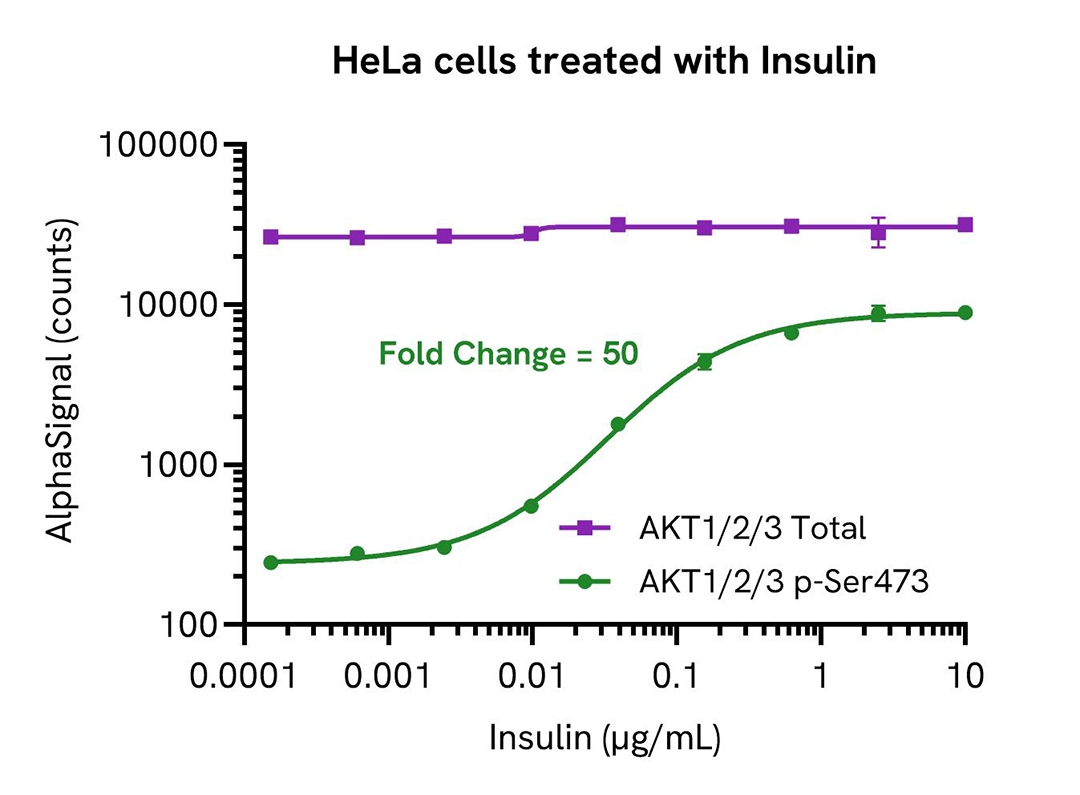
Activation of AKT1/2/3 phospho-(Ser473) in Insulin treated cells
HeLa cells were seeded in a 96-well plate (20,000 cells/well) in complete medium, and incubated overnight at 37°C, 5% CO2. The cells were serum starved for 3 hours and then treated with increasing concentrations of Insulin for 5 minutes.
After treatment, the cells were lysed with 100 µL of Lysis Buffer for 10 minutes at RT with shaking (350 rpm). AKT1/2/3 Phospho (Ser473) and Total levels were evaluated using respective AlphaLISA SureFire Ultra assays. For the detection step, 10 µL of cell lysate (approximately 2,000 cells) was transferred into a 384-well white OptiPlate, followed by 5 µL of Acceptor mix and incubated for 1 hour at RT. Finally, 5 µL of Donor mix was then added to each well and incubated for 1 hour at RT in the dark. The plate was read on an Envision using standard AlphaLISA settings.
As expected, Insulin triggered an increase in levels of Phospho (Ser473) AKT1/2/3 while Total levels remained unchanged.
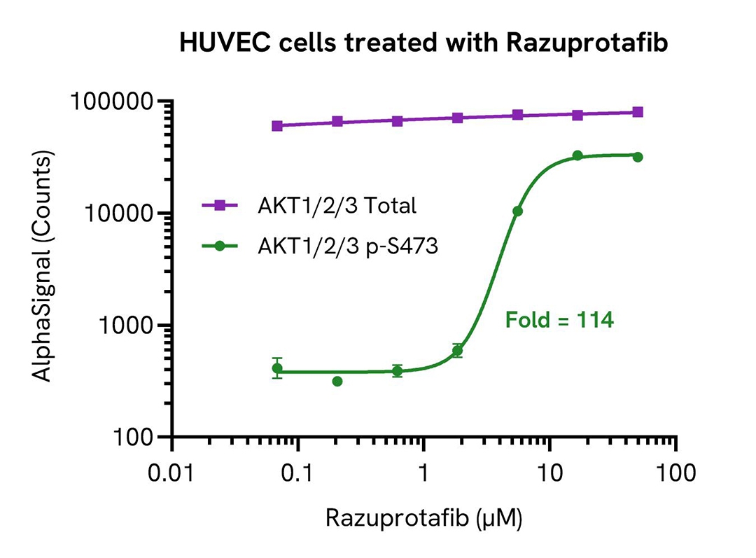
Activation of AKT1/2/3 phospho-(Ser473) in Razuprotafib treated cells
HUVEC cells were seeded in a 96-well plate (20,000 cells/well) in complete medium, and incubated overnight at 37°C, 5% CO2. The cells were serum starved for 2 hours and then treated with increasing concentrations of Razuprotafib for 15 minutes.
After treatment, the cells were lysed with 100 µL of Lysis Buffer for 10 minutes at RT with shaking (350 rpm). AKT1/2/3 Phospho (Ser473) and Total levels were evaluated using respective AlphaLISA SureFire Ultra assays. For the detection step, 10 µL of cell lysate (approximately 2,000 cells) was transferred into a 384-well white OptiPlate, followed by 5 µL of Acceptor mix and incubated for 1 hour at RT. Finally, 5 µL of Donor mix was then added to each well and incubated for 1 hour at RT in the dark. The plate was read on an Envision using standard AlphaLISA settings.
As expected, Razuprotafib triggered an increase in the level of Phospho (Ser473) AKT1/2/3 while Total levels remained unchanged.
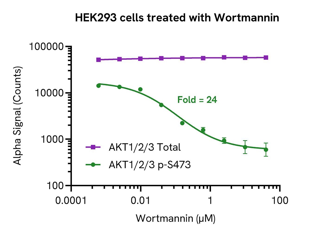
Inhibition of AKT1/2/3 phospho-(Ser473) in Wortmannin treated cells
HEK293 cells were seeded in a 96-well plate (20,000 cells/well) in complete medium, and incubated overnight at 37°C, 5% CO2. The cells were treated with increasing concentrations of Wortmannin for 2 hours.
After treatment, the cells were lysed with 100 µL of Lysis Buffer for 10 minutes at RT with shaking (350 rpm). AKT1/2/3 Phospho (Ser473) and Total levels were evaluated using respective AlphaLISA SureFire Ultra assays. For the detection step, 10 µL of cell lysate (approximately 2,000 cells) was transferred into a 384-well white OptiPlate, followed by 5 µL of Acceptor mix and incubated for 1 hour at RT. Finally, 5 µL of Donor mix was then added to each well and incubated for 1 hour at RT in the dark. The plate was read on an Envision using standard AlphaLISA settings.
As expected, Wortmannin triggered a decrease in the levels of Phospho (Ser473) AKT1/2/3 while Total levels remained unchanged.
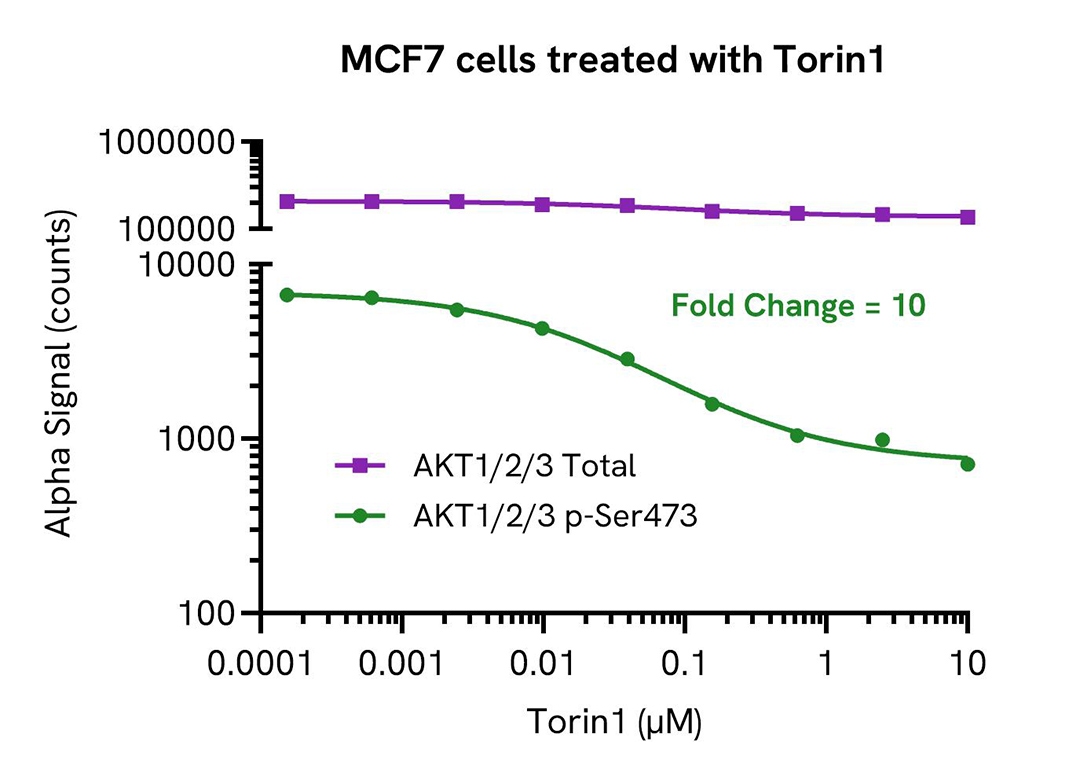
Inhibition of AKT1/2/3 phospho-(Ser473) in Torin1 treated cells
MCF7 cells were seeded in a 96-well plate (40,000 cells/well) in complete medium, and incubated overnight at 37°C, 5% CO2. The cells were treated with increasing concentrations of Torin1 for 18 hours.
After treatment, the cells were lysed with 100 µL of Lysis Buffer for 10 minutes at RT with shaking (350 rpm). AKT1/2/3 Phospho (Ser473) and Total levels were evaluated using respective AlphaLISA SureFire Ultra assays. For the detection step, 10 µL of cell lysate (approximately 4,000 cells) was transferred into a 384-well white OptiPlate, followed by 5 µL of Acceptor mix and incubated for 1 hour at RT. Finally, 5 µL of Donor mix was then added to each well and incubated for 1 hour at RT in the dark. The plate was read on an Envision using standard AlphaLISA settings.
As expected, Torin1 triggered a decrease in the levels of Phospho (Ser473) AKT1/2/3 while Total levels remained unchanged.
Resources
Are you looking for resources, click on the resource type to explore further.
Guide
AlphaLISA SureFire Ultra assay optimization
This guide outlines further possible optimization of cellular and immunoassay parameters to ensure the best possible results are...
Guide
AlphaLISA SureFire Ultra: the ultimate guide for successful experiments
The definitive guide for setting up a successful AlphaLISA SureFire Ultra assay
Several biological processes are regulated by...
Brochure
Alpha SureFire Ultra no-wash immunoassay catalog
Discover Alpha SureFire® Ultra™ assays, the no-wash cellular kinase assays leveraging Revvity's exclusive bead-based technology...
Application Note
Characterizing chemokine receptor inhibitors with AlphaLISA SureFire Ultra, Alpha SureFire Ultra Multiplex and LANCE Ultra cAMP assays
The measurement of protein phosphorylation is a useful tool for measuring the modulation of receptor activation by both antibodies...
Guide
Protein degradation awareness in a single guide
An in-depth review of molecular and cellular pathways
The maintenance of proteostasis, the biological mechanisms that control the...
Flyer
Reagent solutions for autoimmunity research.
Advance your autoimmune disease research and benefit from Revvity broad offering of reagent technologies
SDS, COAs, Manuals and more
Are you looking for technical documents related to the product? We have categorized them in dedicated sections below. Explore now.
Safety data sheet
- 言語English国EU
- 言語English国United States
Certificate of analysis
- Lot NumberU19785Lot DateDecember 6, 2024
- Lot NumberU19856Lot DateDecember 6, 2024
- Lot NumberU19741Lot DateDecember 6, 2024
+ Show next 2
Technical data sheet
- Lot Number-Lot Date-
IFUs, Manuals & more
- Resource TypeManual言語English国-


Related Products


3 of 3
How can we help you?
We are here to answer your questions.

