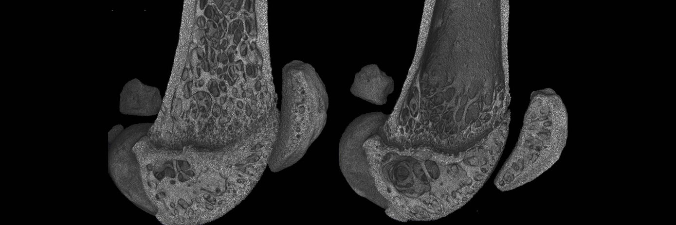

Analysis of bone loss in two osteoporosis mouse models using high resolution microCT imaging
Osteoporosis (OP) is a chronic bone condition that weakens bone mass and density, commonly occurring in post-menopausal women as estrogen levels decrease. There are also acute causes of OP, including corticosteroid-induced OP resulting from extended use of prednisolone (PRED) in immunosuppressive therapies.
MicroCT imaging is a key preclinical imaging modality for bone measurements and often assessments are only done using excised bones at terminal timepoints, requiring high mouse numbers for longitudinal measurements.
This study highlights using the Quantum™ GX3 microCT system to monitor trabecular degeneration and growth plate thickness in two mouse models of bone loss: chronic, ovariectomy-induced (OVEX) OP, and acute PRED associated OP. Both models were assessed at different timepoints to image and measure bone loss in spine and femur. High resolution images were captured both in living animals and harvested bone samples for analyses across the two OP models.
To view the full content please answer a few questions
Download Resource
Analysis of bone loss in two osteoporosis mouse models using high resolution microCT imaging


