メニュー
JP

PhenoVue Fluorescent Probes for Organelles & Subcellular Compartments

Optimized for high-content screening and analysis

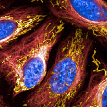
Cellular imaging techniques, such as high-content analysis, rely on the ability to detect and distinguish between specific cellular compartments and organelles. High-quality data depends on high-quality images, which all depends on bright fluorescent dyes. Building on our extensive expertise in imaging instrumentation, fluorescent dye chemistry, and assay development, our PhenoVue organelle-specific stains detect various organelles and cellular compartments and are optimized for high-content analysis and screening. These fluorescent stains can be used for cell membrane, endoplasmic reticulum, golgi apparatus, lipids, lysosomes, nucleoli, nuclei, mitochondria, actin cytoskeleton, and tubulin. For convenience, our multi organelle staining kit contains five ready to use bright fluorescent probes to visualize five key organelles. Applicable to fixed cell models, this kit provides an alternative to the established cell painting assay.

Cell compartment and organelle stains:
  • Cell membrane/endoplasmic reticulum/golgi apparatus: PhenoVue Fluor – WGA and PhenoVue Fluor - Concanavalin A
  • Actin cytoskeleton: PhenoVue Fluor – Phalloidin and PhenoVue Fluor Live Cell Actin Stain
  • Tubulin network: PhenoVue Fluor Live Cell Tubulin Stains
  • Lysosomes: PhenoVue Lysosomal Stains
  • Lipid droplets: PhenoVue Nile Red and PhenoVue 493 Lipid Stain
  • Nucleoli: PhenoVue 512 Nucleic Acid Stain
  • Nuclei: PhenoVue Hoechst 33342 Nuclear Stain, PhenoVue DAPI Nuclear Stain, PhenoVue DRAQ7™ Dead Cell Nuclear Stain, and PhenoVue DRAQ5™ Total Cell Nuclear Stain
  • Mitochondria: PhenoVue Mitochondrial Stains
Features include:
  • Multi organelle staining kit and neuronal differentiation staining kit to streamline your workflow
  • Range of stains and fluors for commonly studied organelles and compartments
  • Variety of fluorescent colors to enable multiplexing while avoiding spectral overlap
  • Validation in high-content screening applications
  • Bright fluorophores to enable high-quality images


For research use only. Not for use in diagnostic procedures.

PhenoVue Fluorescent Probes for Organelles & Subcellular Compartments
bg-element-listing-page bg-element-listing-page-mobile
Products & Services
Resource Library
Filters
Expand All|Collapse All
Application
Brand
Color
Detection Modality
フィルター
Fluorophore
Host Species
Organelle and Cell Compartment
Sample Type
Target Species
タイプ
Sort by: Recommended
Recommended
Product Name A-Z
Product Name Z-A
1 - 25 of 43 Products and Services
PAIBA11 kit primary

PhenoVue™ anti-IBA1 antibody 100X is a monoclonal unconjugated antibody which is part of the PhenoVue Microglia Differentiation Staining Kit. It specifically recognizes IBA1 protein. The expression of IBA1 increases during microglia maturation and is maintained at latest maturation stages. PhenoVue anti-IBA1 antibody is validated for use in imaging and high-content analysis applications.

Part Number: PAIBA11
PMIDIF-11

The PhenoVue™ Microglia Differentiation Staining Kit 1 x 384 comprises a set of validated, optimized and ready-to-use fluorescent probes to vizualize microglia cell in brain cells populations and to monitor microglia differentiation process from progenitors.The kit is validated for use on high-content screening systems such as the Opera Phenix™ Plus high content screening system. 

Part Number: PMIDIF11
PhenoVue Fluor 405LS Phalloidin

PhenoVue™ Fluor 405 – Phalloidin is a fluorescent dye conjugated bicyclic peptide which binds specifically to F-actin and stains the actin cytoskeleton.PhenoVue Fluor 405 conjugated Phalloidin exhibits bright blue fluorescence and can be multiplexed for imaging and high-content screening applications. The PhenoVue Fluor 405 conjugated Phalloidin is validated on Revvity's HCS instruments such as Operetta CLS and Opera Phenix Plus.Part of Revvity's portfolio of cellular imaging reagents, PhenoVue Fluor 405 conjugated Phalloidin has a maximum excitation wavelength of 410 nm and a maximum emission wavelength of 452 nm.

Part Number: CP24051
PhenoVue Fluor 400LS - WGA

PhenoVue Fluor 400LS - WGA is a long Stokes shift fluorescent dye conjugated lectin which displays high affinity for sialic acid and N-acetylglucosamine residues of glycoproteins and glycolipids present at the cellular plasma membranes. It can be used for cellular membrane staining, particularly the Golgi apparatus. Part of Revvity's portfolio of cellular imaging reagents, PhenoVue Fluor 400LS - WGA exhibits bright orange fluorescence and can be easily multiplexed for imaging and high-content screening applications. It has been validated on Revvity's HCS instruments such as Operetta CLS and Opera Phenix Plus, and has a maximum excitation wavelength of 395 nm and a maximum emission wavelength of 585 nm. View our extensive validation data in the Product Information Sheet within the Resources tab below. 

Part Number: CP14001
PhenoVue Fluor 400LS - Concanavalin A

PhenoVue Fluor 400LS - Concanavalin A is a long Stokes shift fluorescent dye conjugated lectin which displays high affinity for glycoproteins and glycolipids present at the cellular membranes. It can be used for cellular membrane staining, particularly the endoplasmic reticulum.Part of Revvity's portfolio of cellular imaging reagents, PhenoVue Fluor 400LS - Concanavalin A exhibits bright orange fluorescence and can be easily multiplexed for imaging and high-content screening applications. It has been validated on Revvity's HCS instruments such as Operetta CLS and Opera Phenix Plus, and has a maximum excitation wavelength of 395 nm and a maximum emission wavelength of 585 nm.View our extensive validation data in the Product Information Sheet within the Resources tab below. 

Part Number: CP94001
PhenoVue Fluor 647 Live Cell Actin Stain - 1x5nmol

PhenoVue™ Fluor 647 live cell actin stain is a no wash, cell permeable fluorogenic dye which specifically bind to actin filaments and is part of Revvity’s portfolio of cellular imaging reagents. PhenoVue Fluor 647 live cell actin stain can be used to visualize actin cytoskeleton in live cells, emiting a far-red fluorescence, and is validated for use in imaging microscopy and high-content screening applications. It exhibits a maximum excitation wavelength at 650 nm and a maximum emission wavelength of 670 nm. View our extensive validation data in the Product Information Sheet within the Resources tab below.

Part Number: CP22R1, CP22R2
PhenoVue anti-MAP2 antibody 100X

PhenoVue anti-MAP2 antibody 100X is a monoclonal unconjugated anti-MAP2 antibody. It specifically recognizes MAP2 (Microtubule-associated protein 2), a neuron-specific protein which promotes the assembly and stability of the microtubule network.MAP2 is essential for the development and the maintenance of neuronal morphology.PhenoVue anti-MAP2 antibody is validated for use in imaging and high-content analysis applications.

Part Number: PABMAP2
PhenoVue Fluor 555 - Phalloidin

PhenoVue™ Fluor 555 - Phalloidin is a fluorescent dye conjugated bicyclic peptide which binds specifically to F-actin and stains the actin cytoskeleton.PhenoVue Fluor 555 conjugated Phalloidin exhibits bright green fluorescence and is validated for use in imaging microscopy and high-content screening applications.Part of Revvity's portfolio of cellular imaging reagents, PhenoVue 555 - Phalloidin has a maximum excitation wavelength of 555 nm and a maximum emission wavelength of 570 nm.View our extensive validation data in the Product Information Sheet within the Resources tab below.

Part Number: CP25551
PhenoVue anti-B3 Tubulin antibody 100X

PhenoVue™ anti-B3 tubulin antibody 100X is a monoclonal unconjugated anti-B3 tubulin antibody which is part of the PhenoVue neuronal differentiation staining kit. It specifically recognizes B3-tubulin, of which expression increases during neuronal maturation and is maintained at latest maturation stages.PhenoVue anti-B3 tubulin antibody is validated for use in imaging and high-content analysis applications. View our extensive validation data in the Product Information Sheet within the Resources tab below.

Part Number: PAB3TUB1
PhenoVue 633 Lysosomal stain

PhenoVue™ 633 lysosomal stain is a fluorescent dye which accumulates in acidic vesicles, such as lysosomes. The bright deep red fluorescence associated with PhenoVue 633 lysosomal stain are validated for use in imaging microscopy and high-content screening systems.Part of Revvity’s portfolio of cellular imaging reagents, the PhenoVue 633 lysosomal stain, has a maximum excitation wavelength of 633 nm and a maximum emission wavelength of 655 nm. View our extensive validation data in the Product Information Sheet within the Resources tab below.

Part Number: CP10DR1
PhenoVue 503 Lysosomal Stain

PhenoVue 503 Lysosomal stain is a fluorescent dye which accumulates in acidic vesicles such as the lysosomes. The bright green fluorescence associated with PhenoVue 503 Lysosomal stain are validated for use in imaging microscopy and High Content Screening Systems.View our extensive validation data in the Product Information Sheet within the Resources tab below.

Part Number: CP10G1
PhenoVue 578 Mitochondrial Stain

PhenoVue 578 Mitochondrial stain is a fluorescent dye which accumulates in heathly mitochondria of live cells.PhenoVue 578 Mitochondria stain exhibits bright red fluorescence and is validated for use in imaging microscopy and high-content screening applications.Part of Revvity's portfolio of cellular imaging reagents, PhenoVue 578 Mitochondrial stain has a maximum excitation wavelength of 578 nm and a maximum emission wavelength of 599 nm, which makes it an alternative to the similar stain MitoTracker™ Red CMTMRos.View our extensive validation data in the Product Information Sheet within the Resources tab below.

Part Number: CP3R1
PhenoVue Nile Red Lipid Stain

PhenoVue Nile Red Lipid stain is a lipophilic organic molecule commonly used for localization and quantification of intracellular lipid droplets. ​ PhenoVue Nile Red Lipid stain exhibits bright yellow fluorescence and is validated for use in imaging microscopy and high-content screening applications. PhenoVue Nile Red Lipid stain exhibits: maximum excitation wavelength of 512nm and maximum emission wavelength of 585 when complexed with triglycerides maximum excitation wavelength of 552nm and maximum emission wavelength of 638nm when complexed with phospholipids View our extensive validation data in the Product Information Sheet within the Resources tab below.

Part Number: CP41
PhenoVue DAPI nuclear stain

PhenoVue™ DAPI nuclear stain is a cell-impermeable organic molecule that binds to DNA and is commonly used to stain cell' nucleus.Part of Revvity’s portfolio of cellular imaging reagents, the PhenoVue DAPI nuclear stain has a maximum excitation wavelength of 348nm and a maximum emission wavelength of 457nm.View our extensive validation data in the Product Information Sheet within the Resources tab below.

Part Number: CP81
PhenoVue Fluor 555 - Concanavalin A

PhenoVue Fluor 555 - Concanavalin A is a fluorescent lectin which displays high affinity for glycoproteins and glycolipids present at the cellular membranes. It can be used for cellular membrane staining, particularly the endoplasmic reticulum. PhenoVue Fluor 555 - Concanavalin A exhibits bright green fluorescence and is validated for use in imaging microscopy and high-content screening applications. Part of Revvity's portfolio of cellular imaging reagents, PhenoVue Fluor 555 - Concanavalin A has a maximum excitation wavelength of 555 nm and a maximum emission wavelength of 570 nm.View our extensive validation data in the Product Information Sheet within the Resources tab below.

Part Number: CP95551
PhenoVue Fluor 647 - Concanavalin A

PhenoVue Fluor 647 - Concanavalin A is a fluorescent lectin which displays high affinity for glycoproteins and glycolipids present at the cellular membranes. It can be used for cellular membrane staining, particularly the endoplasmic reticulum. PhenoVue Fluor 647 - Concanavalin A exhibits bright green fluorescence and is validated for use in imaging microscopy and high-content screening applications. Part of Revvity's portfolio of cellular imaging reagents, PhenoVue Fluor 647 - Concanavalin A has a maximum excitation wavelength of 650 nm and a maximum emission wavelength of 670 nm.View our extensive validation data in the Product Information Sheet within the Resources tab below.

Part Number: CP96471
PhenoVue Fluor 594 - Phalloidin

PhenoVue Fluor 594 - Phalloidin is a fluorescent dye conjugated bicyclic peptide which binds specifically to F-actin and stains the actin cytoskeleton. PhenoVue Fluor 594 conjugated Phalloidin exhibits bright red fluorescence and is validated for use in imaging microscopy and high-content screening applications. Part of Revvity's portfolio of cellular imaging reagents, PhenoVue 594 - Phalloidin has a maximum excitation wavelength of 590 nm and a maximum emission wavelength of 617 nm. View our extensive validation data in the Product Information Sheet within the Resources tab below.

Part Number: CP25941
PhenoVue 551 Mitochondrial Stain

PhenoVue 551 Mitochondrial stain is a fluorescent dye which accumulates in heathly mitochondria of live cells.PhenoVue 551 Mitochondrial stain exhibits bright orange fluorescence and is validated for use in imaging microscopy and high-content screening applications.Part of Revvity's portfolio of cellular imaging reagents, PhenoVue 551 Mitochondrial stain has a maximum excitation wavelength of 551 nm and a maximum emission wavelength of 576 nm, which makes it an alternative to the similar stain MitoTracker™ Orange CMTMRos.View our extensive validation data in the Product Information Sheet within the Resources tab below.

Part Number: CP3O1
PhenoVue Fluor 488 - Concanavalin A

PhenoVue Fluor 488 - Concanavalin A is a fluorescent lectin which displays high affinity for glycoproteins and glycolipids present at the cellular membranes. It can be used for cellular membrane staining, particularly the endoplasmic reticulum.PhenoVue Fluor 488 - Concanavalin A exhibits bright green fluorescence and is validated for use in imaging microscopy and high-content screening applications.Part of Revvity's portfolio of cellular imaging reagents, PhenoVue Fluor 488 - Concanavalin A has a maximum excitation wavelength of 495 nm and a maximum emission wavelength of 520 nm.View our extensive validation data in the Product Information Sheet within the Resources tab below.

Part Number: CP94881
PhenoVue Fluor 488 - WGA

PhenoVue Fluor 488 - WGA is a fluorescent lectin which displays high affinity for sialic acid and N-acetylglucosamine residues of glycoproteins and glycolipids present at the cellular plasma membranes. It can be used for cellular membrane staining, particularly the Golgi apparatus.PhenoVue Fluor 488 - WGA exhibits bright green fluorescence and is validated for use in imaging microscopy and high-content screening applications.Part of Revvity's portfolio of cellular imaging reagents, PhenoVue Fluor 488 - WGA has a maximum excitation wavelength of 495 nm and a maximum emission wavelength of 520 nm.View our extensive validation data in the Product Information Sheet within the Resources tab below.

Part Number: CP14881
PhenoVue Fluor 400LS - Phalloidin

PhenoVue Fluor 400LS - Phalloidin, part of our portfolio of cellular imaging reagents, is a long Stokes shift fluorescent dye conjugated bicyclic peptide which binds specifically to F-actin and stains the actin cytoskeleton.This dye exhibits bright orange fluorescence and can be easily multiplexed for imaging and high-content screening applications. Additionally, it is validated on Revvity' HCS instruments such as Operetta® CLS™ and Opera Phenix® Plus.PhenoVue Fluor 400LS - Phalloidin has a maximum excitation wavelength of 395 nm and a maximum emission wavelength of 585 nm.View our extensive validation data in the Product Information Sheet within the Resources tab below.

Part Number: CP24001
PhenoVue 641 Mitochondrial Stain

PhenoVue 641 Mitochondrial stain is a fluorescent dye which accumulates in heathly mitochondria of live cells. PhenoVue 641 Mitochondria stain exhibits bright red fluorescence and is validated for use in imaging microscopy and high-content screening applications. Part of Revvity's portfolio of cellular imaging reagents, PhenoVue 578 Mitochondrial stain has a maximum excitation wavelength of 641 nm and a maximum emission wavelength of 662 nm, which makes it an alternative to the similar MitoTracker™ Deep Red FM.View our extensive validation data in the Product Information Sheet within the Resources tab below.

Part Number: CP3D1
PhenoVue 493 Lipid Stain

PhenoVue 493 Lipid stain is a lipophilic organic molecule commonly used for localization and quantification of intracellular lipid droplets. ​PhenoVue 493 Lipid stain exhibits bright green fluorescence and is validated for use in imaging microscopy and high-content screening applications. Part of Revvity's portfolio of cellular imaging reagents, PhenoVue 493 Lipid stain has a maximum excitation wavelength of 493 nm and a maximum emission wavelength of 504 nm,which makes it an alternative to the similar stain BODIPY™ 493/503.View our extensive validation data in the Product Information Sheet within the Resources tab below.

Part Number: CP51
MCF-7 cells treated with negative and positive cytotoxic controls for cell death as recorded by DRAQ7

PhenoVue™ DRAQ7™ is a membrane impermeable nuclear stain and is part of Revvity’s portfolio of cellular imaging reagents. It exhibits far-red fluorescence and is validated for use in imaging microscopy and high-content screening applications. PhenoVue DRAQ7 exhibits a maximum excitation wavelength at 599/644 nm and a maximum emission wavelength of 694 nm.View our extensive validation data in the Product Information Sheet within the Resources tab below.

Part Number: CP171
PhenoVue Fluor 594 - WGA

PhenoVue Fluor 594 - WGA is a fluorescent lectin which displays high affinity for sialic acid and N-acetylglucosamine residues of glycoproteins and glycolipids present at the cellular plasma membranes. It can be used for cellular membrane staining, particularly the Golgi apparatus.PhenoVue Fluor 594 - WGA exhibits bright green fluorescence and is validated for use in imaging microscopy and high-content screening applications. Part of Revvity's portfolio of cellular imaging reagents, PhenoVue Fluor 594 - WGA has a maximum excitation wavelength of 590 nm and a maximum emission wavelength of 617 nm.View our extensive validation data in the Product Information Sheet within the Resources tab below.

Part Number: CP15941
Filters
Expand All|Collapse All
Resource Type
Sort by: Resource Name A-Z
Resource Name A-Z
Resource Name Z-A
1 - 25 of 30 Resources
Technical Note
Technical Note
Automated high-content assay using GrowDex-embedded spheroids dispensed on JANUS G3 automated liquid handling workstation

When working with 3D cell models, high heterogeneity between samples can pose a challenge - either biological or technical. In this application note, we demonstrate how a JANUS G3 liquid handling workstation can be used to improve throughput as well as assay statics by automating the liquid handling steps of 384-well, hydrogel-based high-content toxicity assay.

Technical Note
Technical Note
Expanding imaging multiplexing capabilities with PhenoVue Fluor 400LS - Phalloidin

This technical note offers tips and best practices for using PhenoVue ® Fluor 400LS Phalloidin stain to optimize fluorescent signal quality and obtain meaningful biological insights in your research.

Application Note
Application Note
Phenotype discrimination using the PhenoVue cell painting and multi-organelle staining kits

The PhenoVue™ cell painting kit utilizes six fluorescent dyes to reveal a broad range of cellular morphologies and subtle phenotypic changes. But sometimes, a specific research area, cell model, or stage of drug discovery requires a different combination of dyes. For this, Revvity has developed the PhenoVue multi- organelle kit. This kit offers five distinct probes targeting lipid droplets and lysosomes, with a simplified protocol for fixed cells and limited spectral overlap, enabling you to pinpoint the specific cellular structures you’re interested in. In collaboration with the Institute of Bioorganic Chemistry (IBCH) in Poland, we evaluated phenotypic discrimination using 15 reference compounds. Both the PhenoVue cell painting kit and the multi- organelle kit revealed key differences in phenotypic profiles, demonstrating the unique strengths of each assay. Download our app note to learn more.

Product Info
Product Info
PhenoVue 493 Lipid Stain Product Information Sheet

This is a product information sheet for PhenoVue 493 Lipid Stain Product.

Product Info
Product Info
PhenoVue 512 Nucleic Acid Stain Product Information Sheet

This is a product information sheet for PhenoVue 512 Nucleic Acid Stain.

Product Info
Product Info
PhenoVue anti-HSP60 Antibody Product Information Sheet

This is a product information sheet for PhenoVue anti-HSP60 antibody. View validation data, product information, protocols, and more.

Product Info
Product Info
PhenoVue anti-Iba1 antibody

Product Info
Product Info
PhenoVue anti-LAMP1 antibody Product Information Sheet

This is a product information sheet for PhenoVue anti-LAMP antibody. View validation data, product information, protocols, and more.

Product Info
Product Info
PhenoVue anti-MAP2 Antibody

Product Info
Product Info
PhenoVue anti-Nestin Antibody Product Information Sheet

This is a product information sheet for PhenoVue anti-nestin antibody. View validation data, product information, protocols, and more.

Product Info
Product Info
PhenoVue anti-Tubulin Antibody Product Information Sheet

This is a product information sheet for PhenoVue anti-tubulin antibody. View validation data, product information, protocols, and more.

Brochure
Brochure
PhenoVue Cellular Imaging Reagents Brochure

Flyer
Flyer
PhenoVue Cellular Imaging Reagents Flyer

This flyer describes Revvity's PhenoVue cellular imaging reagents.

Product Info
Product Info
PhenoVue DAPI Nuclear Stain Product Information Sheet

This is the product information sheet for PhenoVue DAPI Nuclear Stain.

Product Info
Product Info
Phenovue DRAQ5, Total Cell Nuclear Stain Product Sheet

This is a product information sheet for Phenovue DRAQ5, Total Cell Nuclear Stain

Datasheet
Datasheet
PhenoVue DRAQ7, Dead Cell Nuclear Stain Product Sheet

This is a product information sheet for PhenoVue DRAQ7, Dead Cell Nuclear Stain

Product Info
Product Info
PhenoVue Fluor - Concanavalin A Conjugates Product Information Sheet

Fluorescent Concanavalin A conjugates represent a method of choice for labelling the cellular membranes of mammalian cells, particularly the endoplasmic reticulum which is glycoprotein-enriched.

Product Info
Product Info
PhenoVue Fluor - Phalloidin Conjugates Product Information Sheet

This is a product information sheet for PhenoVue Fluor - Phalloidin Conjugates

Product Info
Product Info
PhenoVue Fluor - WGA Conjugates Product Information Sheet

Fluorescent WGA conjugates represent a method of choice for labelling the cellular membranes of mammalian cells, particularly Golgi apparatus which is glycoprotein enriched.

Product Info
Product Info
PhenoVue Fluor Live Cell Actin Stain

This is a product information sheet for PhenoVue Fluor live cell actin stain. View validation data, product information, protocols, and more.

Product Info
Product Info
PhenoVue Fluor Live Cell Tubulin Stains Product Information Sheet

This is a product information sheet for PhenoVue Fluor Live Cell Tubulin Stains.

Product Info
Product Info
PhenoVue Hoechst 33342 Nuclear Stain Product Information Sheet

This is a product information sheet for PhenoVue Hoechst 33342 Nuclear Stain.

Product Info
Product Info
PhenoVue Lysosomal Stains Product Information Sheet

In living cells, PhenoVue lysosomal stains accumulate in acidic vesicles such as lysosomes, resulting in bright and specific lysosomal fluorescent staining.

Product Info
Product Info
PhenoVue Microglia Differentiation Staining Kit

Product Info
Product Info
PhenoVue Mitochondrial Stains Product Information Sheet

This is a product information sheet for PhenoVue Mitochondrial Stains.