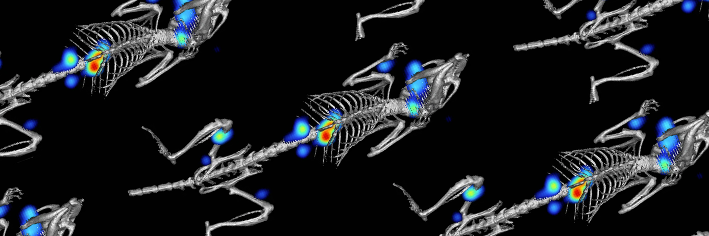
The emergence of non-invasive in vivo imaging technologies has revolutionized our ability to longitudinally track, monitor, and visualize disease progression and treatment effects within a single living organism. Using this approach, researchers can study biological processes in real-time, uncovering insights that were previously unobtainable.
Historically, researchers relied heavily on invasive procedures, often involving the sacrifice of the animals, to gain insights into biological processes and disease states. However, this approach faced significant challenges, including time constraints, high costs, and ethical concerns. It was also difficult to accurately pinpoint specific stages of disease progression or monitor treatment effects when only a single timepoint was being observed.
By contrast, the development of non-invasive in vivo imaging techniques—such as optical, micro CT, and ultrasound—provides researchers with real-time insights about disease mechanisms and therapeutic responses without the need the need for invasive interventions. From early screening to safety evaluation, and from pharmacokinetics to gene therapy, these modalities serve as valuable assets in drug development workflows.
Common applications of in vivo imaging include:
- Early screening to assess drug distribution in the body
- Efficacy assessment to monitor the therapeutic effect of a drug candidate
- Safety evaluation for detecting potential toxicities or adverse effects
- Biodistribution studies to determine the precise location of a drug within a body
- Pharmacokinetics to study how drugs are metabolized, distributed, and eliminated from the body
- Pharmacodynamics to monitor how a drug interacts with its target on a molecular level
- Gene therapy to help tailor drugs to individual patients.
All of these applications yield valuable insights that can be used to inform critical decision-making processes surrounding the safety and efficacy of therapeutic interventions.
The importance of longitudinal imaging in drug discovery
Obtaining longitudinal data through repeated or continuous imaging is important for several reasons, particularly in understanding how diseases progress and the effectiveness of novel treatments. By monitoring biological processes over time, researchers can detect changes in tissue morphology or functional parameters driving disease progression that may not have been evident from single timepoint assessments. It can also determine the effectiveness of therapeutic interventions by tracking changes in disease characteristics, such as tumor size or metabolic activity, over time. This continuous assessment not only allows the identification of early indicators of treatment success or failure, but it can also be used to optimize treatment strategies.
For instance, fluorescence molecular imaging can be used to detect subtle changes at the molecular level, enabling researchers to predict disease progression and therapeutic response long before visible symptoms manifest. Meanwhile, bioluminescence imaging is particularly useful in oncology and infectious disease studies, where it offers a high signal-to-noise ratio simplifying interpretation. Through the use of luminescent reagents, researchers can effectively monitor tumor progression, track pathogen dissemination, and evaluate treatment responses.
Leveraging multimodal imaging for deeper understanding
It is also possible to integrate multiple imaging modalities into a single experiment to gain a more holistic understanding of biological processes within living organisms. For example, optical imaging modalities such as bioluminescence and fluorescence provide high sensitivity for tracking molecular processes and gene expression in vivo. In contrast, ultrasound enables real-time imaging of physiological functions such as blood flow and tissue elasticity, while micro CT offers high spatial resolution for visualizing anatomical structures with fine detail. By leveraging the synergies of these modalities, researchers can obtain a wider range of information that enhances the overall understanding of disease progression, treatment response, and therapeutic efficacy.
This video, presented by Wendy Harp, the product manager for Revvity's in vivo Imaging Portfolio, covers the applications and capabilities of in vivo imaging in drug discovery.

At Revvity, we offer a range of highly sensitive in vivo imaging platforms that provide quality results when paired with Revvity in vivo reagents. To find out more about our in vivo imaging solutions, watch our Understanding Biology webinar series which delves deeper into our capabilities and offerings.
For research use only. Not for use in diagnostic procedures.


