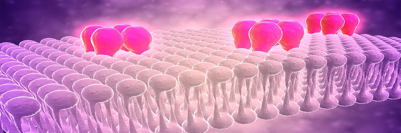
In the early 1970s, Professor Solomon Snyder and colleagues reported a technique that enabled the identification of opiate receptors in the brain.1 This was the first discovery of its kind, providing direct evidence for drug receptors as molecular entities.
Using radioactive ligand binding methods, Dr. Snyder’s team found that the receptors were highly concentrated in areas of the brain involved in the sorts of pain relieved by opiates. They also noted presence of the receptors in nuclei that mediated other pharmacologic actions of opiates, including euphoria, pupillary constriction, and respiratory depression.
This laid the foundations for the future of radioligand binding assays.
Next steps
Following their initial discovery, Snyder’s team extended the technique and identified many major neurotransmitter receptors in the brain, including dopamine receptors, serotonin receptors and α-adrenergic receptors.
In the years that followed, radioligand binding assays became a powerful tool in drug discovery. They can be used to study receptor dynamics and localization, signaling pathways, and compound interactions. The technique is widely used in screening efforts to identify and characterize novel chemical ligands that either block, hinder, or mimic the activity of endogenous chemical messengers with cellular receptors.
Ligands can be divided into two major categories – agonists and antagonists – depending on the effect they have on the receptor. Agonists are molecular entities that bind to and activate a receptor, triggering a biological response. They can either be full or partial agonists, depending on whether they initiate a maximal or submaximal response. One example of a full agonist drug is isoproterenol, which mimics the action of adrenaline at β-adrenoceptors and is used to treat bradycardia conditions.
In contrast, antagonists bind to a target receptor but do not activate it. Instead, they block the effects of competing agonist ligands. Beta blockers are great examples of antagonists, as they act to block the binding of norepinephrine and epinephrine to β-adrenoceptors. Beta blockers are widely used to treat hypertension, angina, and cardiac arrhythmias.
Major types of radioligand studies
As mentioned above, radioligand binding assays can be used to measure the extent or rate of radioligand–receptor binding. Typically, there are three types of assays: saturation, competition, and kinetic experiments. Saturation binding experiments provide information on receptor expression level (Bmax) and the binding affinity of the radiolabeled ligand (KD). Competitive binding experiments determine the concentration of the unlabeled drug that produces radioligand binding halfway between the upper and lower limits, known as the IC50 (inhibitory concentration 50%) or EC50 (effective concentration 50%).
Unlike saturation and competitive binding assays, kinetic binding assays measure the level of binding at various time points. This information is then used to determine the rate constants for radioligand association (Kon) and dissociation (Koff).
Factors to consider when choosing a radioligand
An important consideration is the choice of radioligand for the experiment at hand. Ideally, the radioligand should have high specific activity (>20 Ci/mmol for a tritiated ligand) and low non-specific binding. They should also demonstrate a high degree of selectivity, have a radiochemical purity above 90%, and be stable.
For tips on choosing the right radiochemical for your assay, assay formats, and general information relating to radiometric assays, visit out assay support knowledgebase.
Reference:
- Pert CB, Snyder SH. Opiate receptor: Demonstration in nervous tissue. Science. 1973;179(4077):1011–4.


